

Carotid canal: Difference between revisions
mNo edit summary |
Replaced VE ref names using RefRenamer |
||
| (50 intermediate revisions by 31 users not shown) | |||
| Line 1: | Line 1: | ||
{{Infobox |
{{Infobox bone |
||
| Name = Carotid canal |
|||
| Latin = canalis caroticus |
|||
| Image = Gray141.png |
|||
GraySubject = 34 | |
|||
| ⚫ | |||
GrayPage = 143 | |
|||
| Image2 = |
|||
| ⚫ | |||
| ⚫ | |||
|PartOf=[[Temporal bone]]|System=[[Skeletal system|Skeletal]]}} |
|||
Image2 = | |
|||
The '''carotid canal''' is a passage in the [[petrous part of the temporal bone|petrous part]] of the [[temporal bone]] of the [[skull]] through which the [[internal carotid artery]] and its [[Internal carotid plexus|internal carotid (nervous) plexus]] pass from the neck into (the [[middle cranial fossa]] of) the [[cranial cavity]]. |
|||
| ⚫ | |||
System = | |
|||
MeshName = | |
|||
MeshNumber = | |
|||
DorlandsPre = c_04 | |
|||
DorlandsSuf = 12208545 | |
|||
}} |
|||
The '''carotid canal''' is the passage way in the [[temporal bone]] through which the [[internal carotid artery]] enters the [[middle cranial fossa]] from the neck. The canal starts on the inferior surface of the [[temporal bone]] at the external opening of the carotid canal (also referred to as the carotid foramen). The canal ascends at first vertically, and then, making a bend, runs horizontally forward and medialward. The canal's internal opening is the [[foramen lacerum]]. |
|||
Observing the trajectory of the canal from exterior to interior, the canal is initially directed vertically before curving anteromedially to reach its internal opening.<ref name="www.academie-medecine.fr">{{Cite web |title=canal carotidien l.m. - Dictionnaire médical de l'Académie de Médecine |url=https://www.academie-medecine.fr/le-dictionnaire/index.php?q=canal+carotidien+l.m. |access-date=2024-06-01 |website=www.academie-medecine.fr}}</ref> |
|||
| ⚫ | |||
It transmits into the [[human cranium|cranium]], the [[internal carotid artery]], and the [[carotid plexus]] of nerves. |
|||
== Anatomy == |
|||
| ⚫ | [[ |
||
The carotid canal has two openings, namely internal and external openings.<ref name="pmid27714731" />{{Secondary source needed|date=June 2024}} |
|||
It is divided in three parts, namely, ascending petrous, transverse petrous, and ascending cavernous parts.<ref name="pmid27714731" />{{Secondary source needed|date=June 2024}} |
|||
| ⚫ | |||
=== Boundaries === |
|||
The carotid canal opens into the [[middle cranial fossa]], at the [[petrous part of the temporal bone]]. Anteriorly, it is limited by posterior margin of the [[greater wing of sphenoid bone]]. Posteromedially, it is limited by [[basilar part of occipital bone]].<ref name="pmid27714731" />{{Secondary source needed|date=June 2024}} |
|||
=== Relations === |
|||
The external opening of carotid canal (Latin: "''apertura externa canalis carotici''") is located upon the inferior aspect of the [[petrous part of the temporal bone]]. It is situated anterior to the [[jugular fossa]] (the two being separated by a ridge upon which the [[tympanic canaliculus]] opens inferiorly),<ref>{{Cite web |title=orifice externe du canal carotidien l.m. - Dictionnaire médical de l'Académie de Médecine |url=https://www.academie-medecine.fr/le-dictionnaire/index.php?q=orifice+externe+du+canal+carotidien+l.m. |access-date=2024-06-01 |website=www.academie-medecine.fr}}</ref> and posterolateral to the foramen lacerum.<ref name="pmid27714731" />{{Secondary source needed|date=June 2024}} |
|||
The internal opening of carotid canal (Latin: "''apertura interna canalis carotici''") opens into the [[middle cranial fossa]] at the [[apex of petrous part of temporal bone]].<ref>{{Cite web |title=orifice interne du canal carotidien l.m. - Dictionnaire médical de l'Académie de Médecine |url=https://www.academie-medecine.fr/le-dictionnaire/index.php?q=orifice+interne+du+canal+carotidien+l.m. |access-date=2024-06-01 |website=www.academie-medecine.fr}}</ref> It is situated lateral to [[foramen lacerum]].<ref name="pmid27714731" />{{Secondary source needed|date=June 2024}} |
|||
Both internal and external openings of the carotid canal lie anterior to the [[jugular foramen]] (which opens into the [[posterior cranial fossa]]).<ref name="pmid27714731">{{cite journal | vauthors = Naidoo N, Lazarus L, Ajayi NO, Satyapal KS | title = An anatomical investigation of the carotid canal | journal = Folia Morphologica | volume = 76 | issue = 2 | pages = 289–294 | date = 2017 | pmid = 27714731 | doi = 10.5603/FM.a2016.0060 | url = | doi-access = free }}</ref><ref>{{cite web |last1=Tosovic |first1=Danijel |title=Carotid canal |url=https://www.kenhub.com/en/library/anatomy/carotid-canal |website=www.kenhub.com |access-date=7 June 2024}}</ref> |
|||
The carotid canal is separated from [[middle ear]] and [[inner ear]] by a thin plate of bone.<ref name="Ryan 2011">{{cite book |last1=Ryan |first1=Stephanie |title=Anatomy for diagnostic imaging |date=2011 |publisher=Elsevier Ltd |isbn=9780702029714 |page=80 |edition=Third |chapter=2}}</ref> |
|||
| ⚫ | |||
The canal transmits [[internal carotid artery]] together with its associated [[Internal carotid plexus|nervous plexus]] and [[Internal carotid venous plexus|venous plexus]].<ref name="www.academie-medecine.fr" /><ref name="pmid27714731" />{{Secondary source needed|date=June 2024}} |
|||
== Clinical significance == |
|||
Any skull fractures that damage the carotid canal can put the [[internal carotid artery]] at risk.<ref name="Houseman-2012">{{Cite book|last1=Houseman|first1=Clifford M.|chapter-url=https://www.sciencedirect.com/science/article/pii/B9781437707014000208|title=Principles of Neurological Surgery|last2=Belverud|first2=Shawn A.|last3=Narayan|first3=Raj K.|publisher=[[Saunders (imprint)|Saunders]]|year=2012|isbn=978-1-4377-0701-4|edition=3rd|pages=325–347|language=en|chapter=20 - Closed Head Injury|doi=10.1016/C2009-0-52989-3}}</ref> [[Angiography]] can be used to ensure that there is no damage, and to aid in treatment if there is.<ref name="Houseman-2012" /> |
|||
==Other animals== |
|||
The carotid canal starts on the inferior surface of the [[temporal bone]] of the [[skull]] at the external opening of the carotid canal (also referred to as the carotid foramen). The canal ascends at first superiorly, and then, making a bend, runs anteromedially. Its internal opening is near the [[foramen lacerum]], above which the internal carotid artery passes on its way anteriorly to the [[cavernous sinus]].<ref name="Kumar-2005">{{Cite book|last1=Kumar|first1=Amarendhra M.|chapter-url=https://www.sciencedirect.com/science/article/pii/B0721601375500040|title=Small Animal Ear Diseases|last2=Roman-Auerhahn|first2=Margo Ruth|date=2005-01-01|publisher=[[Saunders (imprint)|Saunders]]|year=2005|isbn=978-0-7216-0137-3|edition=2nd|pages=1–21|language=en|chapter=1 - Anatomy of the Canine and Feline Ear|doi=10.1016/B0-72-160137-5/50004-0}}</ref> |
|||
The carotid canal allows the [[internal carotid artery]] to pass into the [[human cranium|cranium]],<ref name="Kumar-2005" /><ref>{{Cite book|last1=Maynard|first1=Robert Lewis|chapter-url=https://www.sciencedirect.com/science/article/pii/B9780128118375000071|title=Anatomy and Histology of the Laboratory Rat in Toxicology and Biomedical Research|last2=Downes|first2=Noel|date=2019-01-01|publisher=[[Academic Press]]|year=2019|isbn=978-0-12-811837-5|pages=77–90|language=en|chapter=7 - The Cardiovascular System|doi=10.1016/B978-0-12-811837-5.00007-1}}</ref> as well as the [[Internal carotid plexus|carotid plexus]] traveling on the artery.<ref name="Kumar-2005" /> |
|||
| ⚫ | The carotid plexus contains [[sympathetics]] to the head from the [[superior cervical ganglion]].<ref name="Kumar-2005" /> They have several motor functions: raise the [[eyelid]] ([[superior tarsal muscle]]), dilate [[pupil]] (pupillary dilator muscle), innervate [[sweat glands]] of face and [[scalp]] and constricts [[blood vessels]] in the head. |
||
| ⚫ | |||
<gallery> |
<gallery> |
||
File:Gray191.png|Horizontal section of nasal and orbital cavities. |
|||
File:Gray913.png|Coronal section of right temporal bone. |
|||
File:Slide8qqq.JPG|Carotid canal. |
File:Slide8qqq.JPG|Carotid canal. |
||
</gallery> |
</gallery> |
||
== |
== References == |
||
| ⚫ | |||
* {{RocheLexicon|34257.000-1}} |
|||
| ⚫ | |||
{{Gray's}} |
{{Gray's}} |
||
{{Reflist}} |
|||
== External links == |
|||
| ⚫ | |||
* {{cite web|url=http://www.tk.de/rochelexikon/pics/s34257.000-1.html|title=Anatomy diagram: 34257.000-1|work= Roche Lexicon - illustrated navigator|publisher= Elsevier|archive-url=https://web.archive.org/web/20120722052131/http://www.tk.de/rochelexikon/pics/s34257.000-1.html|archive-date=2012-07-22}} |
|||
| ⚫ | |||
{{Skull}} |
{{Skull}} |
||
{{Foramina of skull}} |
{{Foramina of skull}} |
||
{{Portal bar|Anatomy}} |
|||
{{Authority control}} |
|||
[[Category:Foramina of the skull]] |
[[Category:Foramina of the skull]] |
||
[[Category:Skull]] |
|||
[[Category:Bones of the head and neck]] |
|||
[[ar:نفق سباتي]] |
|||
[[Category:Human head and neck]] |
|||
[[de:Canalis caroticus]] |
|||
[[Category:Otorhinolaryngology]] |
|||
[[hu:Canalis caroticus]] |
|||
[[pl:Kanał tętnicy szyjnej]] |
|||
[[pt:Canal carótico]] |
|||
[[ru:Сонный канал]] |
|||
Latest revision as of 23:19, 20 September 2024
| Carotid canal | |
|---|---|
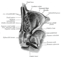
 Left temporal bone. Inferior surface. ("Opening of carotid canal" labeled at center left.) | |
| Details | |
| Part of | Temporal bone |
| System | Skeletal |
| Identifiers | |
| Latin | canalis caroticus |
| TA98 | A02.1.06.013 |
| TA2 | 651 |
| FMA | 55805 |
| Anatomical terms of bone | |
The carotid canal is a passage in the petrous part of the temporal bone of the skull through which the internal carotid artery and its internal carotid (nervous) plexus pass from the neck into (the middle cranial fossa of) the cranial cavity.
Observing the trajectory of the canal from exterior to interior, the canal is initially directed vertically before curving anteromedially to reach its internal opening.[1]
Anatomy
[edit]The carotid canal has two openings, namely internal and external openings.[2][non-primary source needed]
It is divided in three parts, namely, ascending petrous, transverse petrous, and ascending cavernous parts.[2][non-primary source needed]
Boundaries
[edit]The carotid canal opens into the middle cranial fossa, at the petrous part of the temporal bone. Anteriorly, it is limited by posterior margin of the greater wing of sphenoid bone. Posteromedially, it is limited by basilar part of occipital bone.[2][non-primary source needed]
Relations
[edit]The external opening of carotid canal (Latin: "apertura externa canalis carotici") is located upon the inferior aspect of the petrous part of the temporal bone. It is situated anterior to the jugular fossa (the two being separated by a ridge upon which the tympanic canaliculus opens inferiorly),[3] and posterolateral to the foramen lacerum.[2][non-primary source needed]
The internal opening of carotid canal (Latin: "apertura interna canalis carotici") opens into the middle cranial fossa at the apex of petrous part of temporal bone.[4] It is situated lateral to foramen lacerum.[2][non-primary source needed]
Both internal and external openings of the carotid canal lie anterior to the jugular foramen (which opens into the posterior cranial fossa).[2][5]
The carotid canal is separated from middle ear and inner ear by a thin plate of bone.[6]
Contents
[edit]The canal transmits internal carotid artery together with its associated nervous plexus and venous plexus.[1][2][non-primary source needed]
Clinical significance
[edit]Any skull fractures that damage the carotid canal can put the internal carotid artery at risk.[7] Angiography can be used to ensure that there is no damage, and to aid in treatment if there is.[7]
Other animals
[edit]The carotid canal starts on the inferior surface of the temporal bone of the skull at the external opening of the carotid canal (also referred to as the carotid foramen). The canal ascends at first superiorly, and then, making a bend, runs anteromedially. Its internal opening is near the foramen lacerum, above which the internal carotid artery passes on its way anteriorly to the cavernous sinus.[8]
The carotid canal allows the internal carotid artery to pass into the cranium,[8][9] as well as the carotid plexus traveling on the artery.[8]
The carotid plexus contains sympathetics to the head from the superior cervical ganglion.[8] They have several motor functions: raise the eyelid (superior tarsal muscle), dilate pupil (pupillary dilator muscle), innervate sweat glands of face and scalp and constricts blood vessels in the head.
Additional images
[edit]-
 Horizontal section of nasal and orbital cavities.
 -
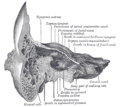 Coronal section of right temporal bone.
 -
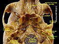 Carotid canal.


References
[edit]![]() This article incorporates text in the public domain from page 143 of the 20th edition of Gray's Anatomy (1918)
This article incorporates text in the public domain from page 143 of the 20th edition of Gray's Anatomy (1918)
- ^ a b "canal carotidien l.m. - Dictionnaire médical de l'Académie de Médecine". www.academie-medecine.fr. Retrieved 2024-06-01.
- ^ a b c d e f g Naidoo N, Lazarus L, Ajayi NO, Satyapal KS (2017). "An anatomical investigation of the carotid canal". Folia Morphologica. 76 (2): 289–294. doi:10.5603/FM.a2016.0060. PMID 27714731.
- ^ "orifice externe du canal carotidien l.m. - Dictionnaire médical de l'Académie de Médecine". www.academie-medecine.fr. Retrieved 2024-06-01.
- ^ "orifice interne du canal carotidien l.m. - Dictionnaire médical de l'Académie de Médecine". www.academie-medecine.fr. Retrieved 2024-06-01.
- ^ Tosovic, Danijel. "Carotid canal". www.kenhub.com. Retrieved 7 June 2024.
- ^ Ryan, Stephanie (2011). "2". Anatomy for diagnostic imaging (Third ed.). Elsevier Ltd. p. 80. ISBN 9780702029714.
- ^ a b Houseman, Clifford M.; Belverud, Shawn A.; Narayan, Raj K. (2012). "20 - Closed Head Injury". Principles of Neurological Surgery (3rd ed.). Saunders. pp. 325–347. doi:10.1016/C2009-0-52989-3. ISBN 978-1-4377-0701-4.
- ^ a b c d Kumar, Amarendhra M.; Roman-Auerhahn, Margo Ruth (2005-01-01). "1 - Anatomy of the Canine and Feline Ear". Small Animal Ear Diseases (2nd ed.). Saunders. pp. 1–21. doi:10.1016/B0-72-160137-5/50004-0. ISBN 978-0-7216-0137-3.
{{cite book}}: CS1 maint: date and year (link) - ^ Maynard, Robert Lewis; Downes, Noel (2019-01-01). "7 - The Cardiovascular System". Anatomy and Histology of the Laboratory Rat in Toxicology and Biomedical Research. Academic Press. pp. 77–90. doi:10.1016/B978-0-12-811837-5.00007-1. ISBN 978-0-12-811837-5.
{{cite book}}: CS1 maint: date and year (link)
External links
[edit]- Atlas image: n3a8p1 at the University of Michigan Health System
- "Anatomy diagram: 34257.000-1". Roche Lexicon - illustrated navigator. Elsevier. Archived from the original on 2012-07-22.
- Photo at Winona.edu