Papers by Monika Liguz-lecznar

Neurobiology of Aging, Oct 1, 2015
Tumor necrosis factor-α (TNF-α) is one of the key players in stroke progression and can interfere... more Tumor necrosis factor-α (TNF-α) is one of the key players in stroke progression and can interfere with brain functioning. We previously documented an impairment of experience-dependent plasticity in the cortex neighboring the stroke-induced lesion, which was accompanied with an upregulation of Tnf-α level in the brain of ischemic mice 1 week after the stroke. Because TNF receptor 1 (TnfR1) signaling is believed to be a major mediator of the cytotoxicity of Tnf-α through activation of caspases, we used an anti-inflammatory intervention aimed at Tnf-α R1 pathway, in order to try to attenuate the detrimental effect of post-stroke inflammation, and investigated if this will be effective in protecting plasticity in the infarct proximity. Aged mice (12-14 months) were subjected to the photothrombotic stroke localized near somatosensory cortex, and immediately after ischemia sensory deprivation was introduced to induce plasticity. Soluble TNF-α R1 (sTNF-α R1), which competed for TNF-α with receptors localized in the brain, was delivered chronically directly into the brain tissue for the whole period of deprivation using ALZET Micro-Osmotic pumps. We have shown that such approach undertaken simultaneously with the stroke reduced the level of TNF-α in the peri-ischemic tissue and was successful in preserving the post-stroke deprivation-induced brain plasticity.
Acta Neurobiologiae Experimentalis, 2019

Acta Neurobiologiae Experimentalis, 2006
Vesicular glutamate transporters are responsible for glutamate transport into synaptic vesicles. ... more Vesicular glutamate transporters are responsible for glutamate transport into synaptic vesicles. In the present study, we examined immunohistochemically the expression of vesicular glutamate transporters, VGluT1 and VGluT2, in trigeminal ganglion neurons of the rat. Immunohistochemistry for VGluT1 and VGluT2 indicated that more than 80% of trigeminal ganglion neurons express VGluT1 and/or VGluT2 in their cell bodies. It also indicated that large and small trigeminal ganglion neurons express VGluT2 more frequently than VGluT1. Dual immunofluorescence histochemistry for VGluT1 and VGluT2 indicated that trigeminal ganglion neurons express VGluT2 more frequently than VGluT1 and that more than 80% of VGluT-expressing trigeminal ganglion neurons express VGluT1 and VGluT2. Many axon terminals in the superficial layers of the medullary dorsal horn also showed VGluT1 and VGluT2 immunoreactivities. Some of these axon terminals were confirmed to form the central core of the synaptic glomerulus. These results indicated that VGluT1 and VGluT2 are coexpressed in the cell bodies and axon terminals in most trigeminal ganglion neurons. J.
ACS Chemical Neuroscience, Sep 26, 2019
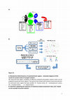
A: Biochemical distinctiveness of examined brain regions -schematic diagram of PCA results for pr... more A: Biochemical distinctiveness of examined brain regions -schematic diagram of PCA results for protein levels of synaptic markers. We observed that higher variability of different analyzed presynaptic markers levels can be assigned to particular brain regions. The name of the marker with variation level characteristic for particular brain region has been bolded. FC-frontal cortex; SC -somatosensory cortex; OC -occipital cortex; H -hippocampus. B: Flowchart explaining the sequence of statistical analysis presented in fig. and based on the example of protein dataset. Table ST1. The design of experiment and analyses. Age groups Young (Y) Middle-aged (A) Old (O) Age of animals 2-3 months old 12-14 months old 22-24 months old Number of mice used in all experiments 12 12 12 Method of analysis Real-time PCR WB HPLC Realtime PCR WB HPLC Realtime PCR WB

Aging Cell, May 12, 2017
As it was established that aging is not associated with massive neuronal loss, as was believed in... more As it was established that aging is not associated with massive neuronal loss, as was believed in the mid-20th Century, scientific interest has addressed the influence of aging on particular neuronal subpopulations and their synaptic contacts, which constitute the substrate for neural plasticity. Inhibitory neurons represent the most complex and diverse group of neurons, showing distinct molecular and physiological characteristics and possessing a compelling ability to control the physiology of neural circuits. This review focuses on the aging of GABAergic neurons and synapses. Understanding how aging affects synapses of particular neuronal subpopulations may help explain the heterogeneity of aging-related effects. We reviewed the literature concerning the effects of aging on the numbers of GABAergic neurons and synapses as well as aging-related alterations in their presynaptic and postsynaptic components. Finally, we discussed the influence of those changes on the plasticity of the GABAergic system, highlighting our results concerning aging in mouse somatosensory cortex and linking them to plasticity impairments and brain disorders. We posit that aging-induced impairments of the GABAergic system lead to an inhibitory/excitatory imbalance, thereby decreasing neuron's ability to respond with plastic changes to environmental and cellular challenges, leaving the brain more vulnerable to cognitive decline and damage by synaptopathic diseases.
Acta Neurobiologiae Experimentalis, 2009
Neurobiology of Aging, Oct 1, 2011
This study addresses the relationship between aging and experience-dependent plasticity in the mo... more This study addresses the relationship between aging and experience-dependent plasticity in the mouse somatosensory cortex. Plasticity in the cortical representation of vibrissae (whiskers) was investigated in young (3 months), mature (14 months) and old (2 years) mice using [14C]2-deoxyglucose (2-DG) autoradiography. Plastic changes were evoked using two experimental paradigms. The deprivation-based protocol included unilateral deprivation of all but one row of whiskers for a week. In the conditioning protocol the animals were subjected to classical conditioning, where tactile stimulation of one row of whiskers was paired with an aversive stimulus. Both procedures evoked functional plasticity in the young group, expressed as a widening of the functional cortical representation of the spared or conditioned row. Aging had a differential effect on these two forms of plasticity. Conditioning-related plasticity was more vulnerable to aging: the plastic change was not detectable in mature animals, even though they acquired the behavioral response. Deprivation-induced plasticity also declined with age, but some effects were persistent in the oldest animals.

International Journal of Developmental Neuroscience, Jan 7, 2007
Vesicular glutamate transporters are responsible for glutamate transport into synaptic vesicles. ... more Vesicular glutamate transporters are responsible for glutamate transport into synaptic vesicles. In the present study, we examined immunohistochemically the expression of vesicular glutamate transporters, VGluT1 and VGluT2, in trigeminal ganglion neurons of the rat. Immunohistochemistry for VGluT1 and VGluT2 indicated that more than 80% of trigeminal ganglion neurons express VGluT1 and/or VGluT2 in their cell bodies. It also indicated that large and small trigeminal ganglion neurons express VGluT2 more frequently than VGluT1. Dual immunofluorescence histochemistry for VGluT1 and VGluT2 indicated that trigeminal ganglion neurons express VGluT2 more frequently than VGluT1 and that more than 80% of VGluT-expressing trigeminal ganglion neurons express VGluT1 and VGluT2. Many axon terminals in the superficial layers of the medullary dorsal horn also showed VGluT1 and VGluT2 immunoreactivities. Some of these axon terminals were confirmed to form the central core of the synaptic glomerulus. These results indicated that VGluT1 and VGluT2 are coexpressed in the cell bodies and axon terminals in most trigeminal ganglion neurons. J.
Frontiers in Neural Circuits, Jun 30, 2016
Since its discovery over four decades ago, somatostatin (SOM) receives growing scientific and cli... more Since its discovery over four decades ago, somatostatin (SOM) receives growing scientific and clinical interest. Being localized in the nervous system in a subset of interneurons somatostatin acts as a neurotransmitter or neuromodulator and its role in the fine-tuning of neuronal activity and involvement in synaptic plasticity and memory formation are widely recognized in the recent literature. Combining transgenic animals with electrophysiological, anatomical and molecular methods allowed to characterize several subpopulations of somatostatin-containing interneurons possessing specific anatomical and physiological features engaged in controlling the output of cortical excitatory neurons. Special characteristic and connectivity of somatostatin-containing neurons set them up as significant players in shaping activity and plasticity of the nervous system. However, somatostatin is not just a marker of particular interneuronal subpopulation. Somatostatin itself acts pre-and postsynaptically, modulating excitability and neuronal responses. In the present review, we combine the knowledge regarding somatostatin and somatostatin-containing interneurons, trying to incorporate it into the current view concerning the role of the somatostatinergic system in cortical plasticity.
Acta Neurobiologiae Experimentalis, 2009

The Journal of Neuroscience, Apr 6, 2011
The somatosensory cortex in mice contains primary (SI) and secondary (SII) areas, differing in so... more The somatosensory cortex in mice contains primary (SI) and secondary (SII) areas, differing in somatotopic precision, topographic organization, and function. The role of SII in somatosensory processing is still poorly understood. SII is activated bilaterally during attentional tasks and is considered to play a role in tactile memory and sensorimotor integration. We measured the plasticity of SII activation after associative learning based on classical conditioning, in which unilateral stimulation of one row of vibrissae was paired with a tail shock. The training consisted of three daily 10 min sessions, during which 40 pairings were delivered. Cortical activation driven by stimulation of vibrissae was mapped with 2-[ 14 C]deoxyglucose (2DG) autoradiography 1 d after the end of conditioning. We reported previously that the conditioning procedure resulted in unilateral enlargement of 2DG-labeled cortical representation of the "trained" row of vibrissae in SI. Here, we measured the width and intensity of the labeled region in SII. We found that both measured parameters in SII increased bilaterally. The increase was observed in cortical layers II/III and IV. Apparently, plasticity in SII is not a simple reflection of changes in SI. It may be attributable to bilateral integrative role of SII, its lesser topographical specificity, and strong involvement in attentional processing.

Pharmacological Reports, Dec 31, 2022
Background Long-term cocaine exposure leads to dysregulation of the reward system and initiates p... more Background Long-term cocaine exposure leads to dysregulation of the reward system and initiates processes that ultimately weaken its rewarding effects. Here, we studied the influence of an escalating-dose cocaine regimen on drug-associated appetitive behavior after a withdrawal period, along with corresponding molecular changes in plasma and the prefrontal cortex (PFC). Methods We applied a 5 day escalating-dose cocaine regimen in rats. We assessed anxiety-like behavior at the beginning of the withdrawal period in the elevated plus maze (EPM) test. The reinforcement properties of cocaine were evaluated in the Conditioned Place Preference (CPP) test along with ultrasonic vocalization (USV) in the appetitive range in a drugassociated context. We assessed corticosterone, proopiomelanocortin (POMC), β-endorphin, CART 55-102 levels in plasma (by ELISA), along with mRNA levels for D2 dopaminergic receptor (D2R), κ-receptor (KOR), orexin 1 receptor (OX1R), CART 55-102, and potential markers of cocaine abuse: miRNA-124 and miRNA-137 levels in the PFC (by PCR). Results Rats subjected to the escalating-dose cocaine binge regimen spent less time in the cocaine-paired compartment, and presented a lower number of appetitive USV episodes. These changes were accompanied by a decrease in corticosterone and CART levels, an increase in POMC and β-endorphin levels in plasma, and an increase in the mRNA for D2R and miRNA-124 levels, but a decrease in the mRNA levels for KOR, OX1R, and CART 55-102 in the PFC. Conclusions The presented data reflect a part of a bigger picture of a multilevel interplay between neurotransmitter systems and neuromodulators underlying processes associated with cocaine abuse. Cocaine binge • HPA axis • Neuromodulators • KOR • OX1R • microRNA Abbreviations ACTH Adrenocorticotropic hormone BDNF Brain-derived neurotrophic factor CART 55-102 Cocaine and amphetamine-regulated transcript 55-102 CB rats Cocaine 'binged' rats CNS Central nervous system CPP test Conditioned place preference test CRF Corticotropin-releasing factor D2R D2 dopaminergic receptor DAT Dopamine active transporter ELISA Enzyme-linked immunosorbent assay EPM test Elevated plus maze test HPA axis Hypothalamic-pituitary-adrenal axis KOR κ-Opioid receptor LV Lentiviral vector mPFC Medial prefrontal cortex Małgorzata Lehner: Deceased

PubMed, 2007
Glutamate is the predominant excitatory neurotransmitter in the central nervous system (CNS) and ... more Glutamate is the predominant excitatory neurotransmitter in the central nervous system (CNS) and glutamatergic transmission is critical for controlling neuronal activity. Glutamate is stored in synaptic vesicles and released upon stimulation. The homeostasis of glutamatergic system is maintained by a set of transporters present in plasma membrane and in the membrane of synaptic vesicles. The family of vesicular glutamate transporters in mammals is comprised of three highly homologous proteins: VGLUT1-3. The expression of particular VGLUTs is largely complementary with limited overlap and so far they are most specific markers for neurons that use glutamate as neurotransmitter. VGLUTs are regulated developmentally and determine functionally distinct populations of glutamatergic neurons. Controlling the activity of these proteins could potentially modulate the efficiency of excitatory neurotransmission. This review summarizes the recent knowledge concerning molecular and functional characteristic of vesicular glutamate transporters, their development, contribution to synaptic plasticity and their involvement in pathology of the nervous system.
Scientific Reports, Oct 26, 2022
The activity of inhibitory interneurons has a profound role in shaping cortical plasticity. Somat... more The activity of inhibitory interneurons has a profound role in shaping cortical plasticity. Somatostatinexpressing interneurons (SOM-INs) are involved in several aspects of experience-dependent cortical rewiring. We addressed the question of the barrel cortex SOM-IN engagement in plasticity formation induced by sensory deprivation in adult mice (2-3 months old). We used a spared vibrissa paradigm, resulting in a massive sensory map reorganization. Using chemogenetic manipulation, the activity of barrel cortex SOM-INs was blocked or activated by continuous clozapine N-oxide (CNO) administration during one-week-long deprivation. To visualize the deprivation-induced plasticity, [ 14 C]-2-deoxyglucose mapping of cortical functional representation of the spared whisker was performed at the end of the deprivation. The plasticity was manifested as an extension of cortical activation in response to spared vibrissae stimulation. We found that SOM-IN inhibition in the cortical column of the spared whisker did not influence the areal extent of the cortex activated by the spared whisker. However, blocking the activity of SOM-INs in the deprived column, adjacent to the spared one, decreased the plasticity of the spared whisker representation. SOM-IN activation did not affect plasticity. These data show that SOM-IN activity is part of cortical circuitry that affects interbarrel interactions underlying deprivation-induced plasticity in adult mice.

Biochimica et biophysica acta. Molecular cell research, 2022
Gaba-ergic neurons are a diverse cell class with extensive influence over cortical processing, bu... more Gaba-ergic neurons are a diverse cell class with extensive influence over cortical processing, but their role in experience-dependent plasticity is not completely understood. Here we addressed the role of cortical somatostatin- (SOM-INs) and vasoactive intestinal polypeptide- (VIP-INs) containing interneurons in a Pavlovian conditioning where stimulation of the vibrissae is used as a conditioned stimulus and tail shock as unconditioned one. This procedure induces a plastic change observed as an enlargement of the cortical functional representation of vibrissae activated during conditioning. Using layer-targeted, cell-selective DREADD transductions, we examined the involvement of SOM-INs and VIP-INs activity in learning-related plastic changes. Under optical recordings, we injected DREADD-expressing vectors into layer IV (L4) barrels or layer II/III (L2/3) areas corresponding to the activated vibrissae. The activity of the interneurons was modulated during all conditioning sessions, and functional 2-deoxyglucose (2DG) maps were obtained 24 h after the last session. In mice with L4 but not L2/3 SOM-INs suppressed during conditioning, the plastic change of whisker representation was absent. The behavioral effect of conditioning was disturbed. Both L4 SOM-INs excitation and L2/3 VIP-INs inhibition during conditioning did not affect the plasticity or the conditioned response. We found the activity of L4 SOM-INs is indispensable in the formation of learning-induced plastic change. We propose that L4 SOM-INs may provide disinhibition by blocking L4 parvalbumin interneurons, allowing a flow of information into upper cortical layers during learning.

Postepy biochemii, Dec 29, 2018
S omatostatin is a peptide that participates in numerous biochemical and signaling path- ways. It... more S omatostatin is a peptide that participates in numerous biochemical and signaling path- ways. It functions via receptors (SSTRs1-5), which belong to the family of receptors coupled with protein G. All somatostatin receptors are characterized by a certain degree of homology in molecular structure. The cell effects of their agonists in peripheral tissues rely mainly on the inhibition of the hormones release. Somatostatin is also an important neuromodulator and neurotransmitter. SSTRs may affect other receptors, forming structural and functional homodimers and heterodimers. SSTRs play also role in the regulation of physiological processes, such as itching and pain, reproductive functions, regulation of feeding or mood. Besides physiological functions, SSTRs contribute also to the pathogenesis of glial tumors, neurodegenerative diseases, or post hemorrhagic stroke changes. Recent years of research have provided new data regarding the role of somatostatin receptor signaling pathways in the brain and the knowledge in this field is developing rapidly.

PLOS ONE, Dec 7, 2015
Experience-induced plastic changes in the cerebral cortex are accompanied by alterations in excit... more Experience-induced plastic changes in the cerebral cortex are accompanied by alterations in excitatory and inhibitory transmission. Increased excitatory drive, necessary for plasticity, precedes the occurrence of plastic change, while decreased inhibitory signaling often facilitates plasticity. However, an increase of inhibitory interactions was noted in some instances of experience-dependent changes. We previously reported an increase in the number of inhibitory markers in the barrel cortex of mice after fear conditioning engaging vibrissae, observed concurrently with enlargement of the cortical representational area of the row of vibrissae receiving conditioned stimulus (CS). We also observed that an increase of GABA level accompanied the conditioning. Here, to find whether unaltered GABAergic signaling is necessary for learning-dependent rewiring in the murine barrel cortex, we locally decreased GABA production in the barrel cortex or reduced transmission through GABAA receptors (GABAARs) at the time of the conditioning. Injections of 3-mercaptopropionic acid (3-MPA), an inhibitor of glutamic acid decarboxylase (GAD), into the barrel cortex prevented learninginduced enlargement of the conditioned vibrissae representation. A similar effect was observed after injection of gabazine, an antagonist of GABAARs. At the behavioral level, consistent conditioned response (cessation of head movements in response to CS) was impaired. These results show that appropriate functioning of the GABAergic system is required for both manifestation of functional cortical representation plasticity and for the development of a conditioned response.
Biomolecules, Feb 15, 2022
Despite the obvious differences in the pathophysiology of distinct neuropsychiatric diseases or n... more Despite the obvious differences in the pathophysiology of distinct neuropsychiatric diseases or neurodegenerative disorders, some of them share some general but pivotal mechanisms, one of which is the disruption of excitation/inhibition balance. Such an imbalance can be generated by changes in the inhibitory system, very often mediated by somatostatin-containing interneurons (SOM-INs). In physiology, this group of inhibitory interneurons, as well as somatostatin itself, profoundly shapes the brain activity, thus influencing the behavior and plasticity; however, the changes in the number, density and activity of SOM-INs or levels of somatostatin are found throughout many neuropsychiatric and neurological conditions, both in patients and animal models. Here, we (1) briefly describe the brain somatostatinergic system, characterizing the neuropeptide somatostatin itself, its receptors and functions, as well the physiology and circuitry of SOM-INs; and (2) summarize the effects of the activity of somatostatin and SOM-INs in both physiological brain processes and pathological brain conditions, focusing primarily on learning-induced plasticity and encompassing selected neuropsychological and neurodegenerative disorders, respectively. The presented data indicate the somatostatinergic-system-mediated inhibition as a substantial factor in the mechanisms of neuroplasticity, often disrupted in a plethora of brain pathologies.
Brain Structure & Function, Dec 23, 2019
Inhibitory interneurons in the cerebral cortex contain specific proteins or peptides characterist... more Inhibitory interneurons in the cerebral cortex contain specific proteins or peptides characteristic for a certain interneuron subtype. In mice, three biochemical markers constitute non-overlapping interneuron populations, which account for 80-90% of all inhibitory cells. These interneurons express parvalbumin (PV), somatostatin (SST), or vasoactive intestinal peptide (VIP). SST is not only a marker of a specific interneuron subtype, but also an important neuropeptide that participates in numerous biochemical and signalling pathways in the brain via somatostatin receptors (SSTR1-5). In the nervous system, SST acts as a neuromodulator and neurotransmitter affecting, among others, memory, learning, and mood. In the sensory cortex, the co-localisation of GABA and SST is found in approximately 30% of interneurons. Considering the importance of interactions between inhibitory interneurons in cortical plasticity and the possible GABA and SST co-release, it seems important to investigate the localisation of different SSTRs on cortical interneurons. Here, we examined the distribution of SSTR1-5 on barrel cortex interneurons containing PV, SST, or VIP. Immunofluorescent staining using specific antibodies was performed on brain sections from transgenic mice that expressed red fluorescence in one specific interneuron subtype (PV-Ai14, SST-Ai14, and VIP-Ai14 mice). SSTRs expression on PV, SST, and VIP interneurons varied among the cortical layers and we found two patterns of SSTRs distribution in L4 of barrel cortex. We also demonstrated that, in contrast to other interneurons, PV cells did not express SSTR2, but expressed other SSTRs. SST interneurons, which were not found to make chemical synapses among themselves, expressed all five SSTR subtypes.

Progress in Neuro-Psychopharmacology and Biological Psychiatry, 2019
The aim of this study was to assess the influence of chronic restraint stress on amphetamine (AMP... more The aim of this study was to assess the influence of chronic restraint stress on amphetamine (AMPH)related appetitive 50-kHz ultrasonic vocalisations (USVs) in rats differing in freezing duration in a contextual fear test (CFT), i.e. HR (high-anxiety responsive) and LR (low-anxiety responsive) rats. The LR and the HR rats, previously exposed to an AMPH binge experience, differed in sensitivity to AMPH's rewarding effects, measured as appetitive vocalisations. Moreover, chronic restraint stress attenuated AMPH-related appetitive vocalisations in the LR rats but had no influence on the HR rats' behaviour. To specify, the restraint LR rats vocalised appetitively less in the AMPH-associated context and after an AMPH challenge than the control LR rats. This phenomenon was associated with a decrease in the mRNA level for D2 dopamine receptor in the amygdala and its protein expression in the basal amygdala (BA) and opposite changes in the nucleus accumbens (NAc) -an increase in the mRNA level for D2 dopamine receptor and its protein expression in the NAc shell, compared to control conditions. Moreover, we observed that chronic restraint stress influenced epigenetic regulation in the LR and the HR rats differently. The contrasting changes were observed in the dentate gyrus (DG) of the hippocampus -the LR rats presented a decrease, but the HR rats showed an increase in H3K9 trimethylation. The restraint LR rats also showed higher miR-494 and miR-34c levels in the NAc than the control LR group. Our study provides behavioural and biochemical data concerning the role of differences in fear-conditioned response in stress vulnerability and AMPHassociated appetitive behaviour. The LR rats were less sensitive to the rewarding effects of AMPH when previously exposed to chronic stress that was accompanied by changes in D2 dopamine receptor expression and epigenetic regulation in mesolimbic areas. Binged low (LR) and high (HR) anxiety rats differ in AMPH-associated vocalisations Chronic stress dampens appetitive AMPH-associated vocalisations only in LR rats Stress and AMPH binge increase D2 receptor expression in nucleus accumbens in LR rats Stress and AMPH binge decrease D2 receptor expression in amygdala in LR rats Binged stressed LR rats show increased miRNA-34c and miRNA-494 in nucleus accumbens


Uploads
Papers by Monika Liguz-lecznar