Papers by Yasuhiro Takeshima

Tetrasomy 9p is a rare clinical syndrome and about 30% of known cases exhibit chromosome mosaicis... more Tetrasomy 9p is a rare clinical syndrome and about 30% of known cases exhibit chromosome mosaicism. The cases with tetrasomy 9p mosaicism have been reported to show the various phenotypes. On the other hand, Klinefelter syndrome is well recognized chromosomal abnormality caused by an additional X chromosome in males (47,XXY), and the characteristic clinical findings include tall stature, immaturity of external genitalia, testicular dysfunction. Here, we report a 10-year-old male with tetrasomy of 9p mosaicism, whose phenotypic feature is mimicking Klinefelter syndrome. He was referred to our hospital for inconspicuous penis. He showed tall height (+2.5 SD). Endocrinological examination revealed the poor testosterone response to human chorionic gonadotropin administration, which indicated the testicular hypofunction, whereas MRI revealed concealed penis as a cause of inconspicuous penis. Because of the phenotype mimicking Klinefelter syndrome, karyotype of his blood lymphocytes was analyzed, and an additional marker chromosome was detected in 6% of the investigated metaphases. Fluorescence in situ hybridization analysis revealed that the marker chromosome was an isochromosome 9p, which resulted in tetrasomy 9p. Chromosome analysis of buccal smear also showed mosaicism for two karyotypes: 5% of cells had the isochromosome of 9p, and the other cells showed normal. This case is the second case with tetrasomy 9p mosaicism mimicking Klinefelter syndrome phenotype in the world. Our case, together with previously reported cases with the same association, indicates the possibility of testicular hypofunction and urogenital anomalies induced by overexpression of some genes on chromosome 9p.

Most patients with spinal muscular atrophy (SMA) have been reported to show homozygous deletion o... more Most patients with spinal muscular atrophy (SMA) have been reported to show homozygous deletion of the gene responsible for SMA, SMN1. However, whether SMA patients with homozygous deletion of the gene exist in Southeast Asian countries, including Vietnam, remains to be determined, because molecular genetic analyses of SMA patients from these countries have not been reported. In this preliminary study, we analyzed five Vietnamese SMA patients and found that SMN1 gene exons 7 and 8 were completely absent in one of them, a 6-month-old girl with hypotonic muscles. Thus, homozygous deletion of the gene can be a cause of SMA in Vietnam, although other genetic abnormalities should be considered as etiological factors in many cases. In conclusion, we identified a homozygous deletion of the SMN1 gene in a Vietnamese SMA patient. Since the number of the patients analyzed in this study was very limited, it is too early to determine whether homozygous deletion of the gene is not a main cause of SMA in Vietnam.
No to hattatsu = Brain and development, 2004
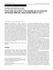
Ornithine transcarbamylase (OTC) deficiency is the most common inborn error of the urea cycle. Al... more Ornithine transcarbamylase (OTC) deficiency is the most common inborn error of the urea cycle. Although a combination of molecular methods have been used including DNA sequencing of all 10 exons and exon-intron boundaries of OTC gene, only ~80% of patients with OTC deficiency are found to have mutations. We report two known and three novel mutations of the OTC gene in five Japanese patients including two neonatal-onset, one late-onset, and two symptomatic female patients. Known nonsense mutations (c.578G>A and c.421C>T) were detected in a neonatal-onset male and a symptomatic female patient, respectively. Mutation analysis revealed two novel mutations including one splice site mutation (c.386+1G>C) in a symptomatic female patient and one missense mutation (c.515T>A) in a late-onset male patient. In the remaining case, which was a neonatal-onset male patient, no mutation was disclosed by direct sequencing of all 10 exons and their flanking intron sequences. Therefore, OTC mRNA in the liver was analyzed by RT-PCR, and remarkably, a 135-nt insertion was detected between exons 5 and 6. Genomic DNA analysis of intron sequences revealed a single nucleotide change at 265 bp downstream from the 3' end of exon 5, which created the novel splice acceptor site. Thereby, a 135-nt exon was created from the central part of an intron sequence. This is the first report of mutation deep in the intronic sequence in the OTC gene. Molecular analysis using genomic DNA and mRNA will increase the mutation detection ratio in the OTC gene. Ornithine transcarbamylase (OTC) deficiency (OTCD; MIM#311250) is the most common inborn error of the urea cycle. OTC catalyzes the formation of citrulline from carbamyl phosphate and ornithine. The human OTC gene is located on the short arm of the X chromosome within band Xp21.1 . The gene spans 73kb with an open reading frame of 1,062 nucleotides and contains 10 exons . The OTC gene is expressed exclusively in the liver and small intestinal mucosa. It encodes a precursor OTC protein containing 354 amino acids and the amino terminus contains a leader sequence of 32 amino acids which is cleaved in two steps upon incorporation into the mitochondrial matrix .
Journal of human genetics, 2002
The dystrophin gene, which is mutated in Duchenne muscular dystrophy, is the largest human gene. ... more The dystrophin gene, which is mutated in Duchenne muscular dystrophy, is the largest human gene. A full spectrum of the gene transcripts has not been fully elucidated yet, although two cryptic exons have so far been identified in the 5' region of the dystrophin gene. Here, a novel dystrophin mRNA containing a 62-nucleotide insertion between exons 3 and 4 was identified in lymphocytes from a Japanese Duchenne muscular dystrophy patient with a single nucleotide deletion in exon 5. The inserted 62-nucleotide sequence was found to be homologous to part of intron 3 and it was revealed that the insertion possessed branch point and both acceptor and donor splice site consensus sequences perfectly. Therefore, the 62-bp insertion sequence was considered to be a novel exon and was designated as exon 3a. However, this insertion was not present in the patient's muscle and 12 different normal tissues that were screened. The physiological role of the novel cryptic exon remains to be clari...

Human genetics, 2003
Intron 2 of the dystrophin gene is unusually large, extending 157 kb on the X-chromosome, and is ... more Intron 2 of the dystrophin gene is unusually large, extending 157 kb on the X-chromosome, and is known to contain one cryptic exon 2a. Here, we report that a single nucleotide change in the middle of this huge intron is a source of two novel extra exons. A novel point mutation changing T to A nucleotide was identified at 5591 bp downstream from the 3' end of exon 2 (T310+5591A) in genomic DNA of an asymptomatic dystrophinopathy case. The mutation identification was initiated by detection of two novel dystrophin mRNAs containing a 132-nucleotide or 46-nucleotide insertion between exons 2 and 3 in lymphocytes but one with a 132-nucleotide insertion in skeletal muscle. It was concluded that T310+5591A created a novel consensus sequence for a splice acceptor site leading to the formation of two novel exon structures by using two cryptic splice donor sites at 132 bp or 46 bp downstream. The former maintained the dystrophin reading frame and was expected to insert 44 amino acids in th...
No to hattatsu. Brain and development, 2014
To examine indications for the early diagnosis of Menkes disease (MD). We compared clinical exami... more To examine indications for the early diagnosis of Menkes disease (MD). We compared clinical examination results of 3 neonate males with family histories of MD who were at risk of being affected (1 infant was affected and 2 were unaffected). Kinky hair, a characteristic shown by MD patients, was observed just after birth in the affected infant; however, this characteristic was extremely mild and easy to miss. In the first month after birth, serum copper level declined over time in the affected infant,while it increased in the unaffected infants. Urine homovanillic acid/vanillylmandelic acid (HVA/VMA) ratio, which is typically high in MD patients, was observed to be 4.9-8.0 (cut-off, 4.0) in the affected infant. In the unaffected infants, the urine HVA/MVA ratio showed a high value of 11.0 during catecholamine administration, but this decreased to below the cut-off value when catecholamine was not administered. Variations in the serum copper level and urine HVA/VMA ratio over time wer...

Pediatrics International, 2009
Background: Congenital generalized lipodystrophy (CGL), Berardinelli‐Seip syndrome, is a rare au... more Background: Congenital generalized lipodystrophy (CGL), Berardinelli‐Seip syndrome, is a rare autosomal recessive disorder characterized by the generalized absence of adipose tissue at birth, severe insulin resistance early in life, hypertriglyceridemia, hepatomegaly, and the development of diabetes mellitus during puberty. Recently, two genes, BSCL2 and AGPAT2, were identified as causative genes for CGL. It has been reported that patients with BSCL mutations present with more severe clinical findings than those with AGPAT2 mutations. However, the clinical course of CGL caused by BSCL2 mutations in infancy has not been fully elucidated.Methods: Two Japanese infantile patients with CGL from independent families were examined and underwent an oral glucose tolerance test. Insulin resistance and insulin secretion were estimated using the homeostasis model assessment for insulin resistance and the insulinogenic index, respectively. Sequence analysis of the entire coding region of BSCL2...
No to hattatsu. Brain and development, 2014

Pediatrics International, 2008
Background: The SMN1 gene is now recognized as a spinal muscular atrophy (SMA)‐causing gene, whil... more Background: The SMN1 gene is now recognized as a spinal muscular atrophy (SMA)‐causing gene, while SMN2 and NAIP have been characterized as a modifying factor of the clinical severity of SMA. Gene dosage of SMN2 is associated with clinical severity of SMA. But the relationship between gene dosage of NAIP and clinical severity of SMA remains to be clarified, although complete deletion of NAIP is frequent in type I patients.Methods: To evaluate the contribution of the SMN2 and NAIP gene dosages to SMA, quantitative real‐time polymerase chain reaction was used to measure copy numbers of SMN2 and NAIP in 34 Vietnamese SMA patients lacking SMN1 (13 type I, 11 type II and 10 type III patients).Results: The SMN2 copy number in type I patients was significantly lower than that in type II–III patients, which was compatible with the previous reports. In contrast, 25 out of 34 patients had only zero or one copy of NAIP, while 50 out of 52 controls had two or more copies. For NAIP (+) genotype,...

Molecular Genetics and Metabolism, 2010
Congenital generalized lipodystrophy (CGL), characterized by generalized absence of adipose tissu... more Congenital generalized lipodystrophy (CGL), characterized by generalized absence of adipose tissue, has heterogeneous causes. Recently, a novel type of CGL complicated by muscular dystrophy was categorized as CGL4 caused by PTRF-CAVIN deficiency. However, it is unknown whether CGL4 exhibits clinical abnormalities during the infantile period. Here, we describe the youngest Japanese case of CGL4-a Japanese girl with asymptomatic high serum creatine kinase (CK) levels at 3 months old. She was referred to our hospital at 5 months of age because of her elevated serum CK (2528 IU/L). Generalized absence of adipose tissue was first recognized at 2 years of age. Mutation analysis of genes known to be responsible for CGL1-3 failed to disclose any abnormalities. Instead, analysis of the PTRF-CAVIN gene encoding PTRF-CAVIN revealed compound heterozygous mutations, one allele contained an insertion (c.696_697insC) and the other allele harbored a novel nonsense mutation (c.512CNA). Our patient had low serum leptin and adiponectin levels and insulin resistance. Pathological studies on biopsied muscle disclosed mild dystrophic change and highly reduced expression of PTRF-CAVIN. It was concluded that our PTRF-CAVIN deficient patient showed not only CGL but also asymptomatic elevation of serum CK because of her mild muscle dystrophic change.

Journal of Human Genetics, 2010
Non-autonomous retrotransposon-mediated mobilizations of the Alu family are known pathogenic mech... more Non-autonomous retrotransposon-mediated mobilizations of the Alu family are known pathogenic mechanisms of human disease. Here, we report a pathogenic, contemporary, non-autonomous retrotransmobilization of part of a novel non-coding gene into the dystrophin gene. In a Japanese Duchenne muscular dystrophy patient, a 330-bp-long de novo insertion was identified in exon 67 of dystrophin. The insertion induced exon 67-skipping in the dystrophin mRNA, creating a premature stop codon. The sequence of the insertion had certain characteristics of retrotransposons: an antisense polyadenylation signal accompanied by a poly(T) sequence and a target site duplication. The insertion site matched the consensus recognition sequence for the L1 endonuclease, indicating a retrotransposon-mediated event, although the inserted sequence did not match any known retrotransposons. The origin of the inserted sequence was mapped to a gene-poor region of chromosome 11. The inserted fragment was expressed in multiple human tissue RNAs, indicating that it is a novel transcript. The full length of the transcript was cloned and showed no meaningful protein coding ability.
Journal of Biochemistry, 2007
Proper splicing is known to proceed under the control of conserved cis-elements located at exon-i... more Proper splicing is known to proceed under the control of conserved cis-elements located at exon-intron boundaries. Recently, it was shown that additional elements, such as exonic splicing enhancers (ESEs), are essential for the proper splicing of certain exons, in addition to the splice donor and acceptor site sequences; however, the relationship between these cis-elements is still unclear. In this report, we utilize dystrophin exon 19 to analyse the relationship between the ESE and its upstream acceptor site sequences. Dystrophin exon 19, which maintains adequate splicing donor and acceptor consensus sequences, encodes exonic splicing enhancer (dys-ESE19) sequences. Splice pattern analysis, using a minigene reporter expressed in HeLa cells, showed that either a strong polypyrimidine tract (PPT) or a fully active dys-ESE19 is sufficient for proper splicing. Each of these two cis-elements has enough activity for proper exon 19 splicing suggesting that the PPT, which is believed to be an essential cis-element for splicing, is dispensable when the downstream exon contains a strong ESE. This compensation was only seen in living cells but not in 'in vitro splicing'. This suggests the possibility that the previous splicing experiments using an in vitro splicing system could underestimate the activity of ESEs.
Human Molecular Genetics, 1999
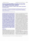
Exon skipping by alternative splicing and circular RNA formation are proposed to be interrelated ... more Exon skipping by alternative splicing and circular RNA formation are proposed to be interrelated events. Since multiple patterns of alternative splicing have been demonstrated in both the 5′ and 3′ regions of the dystrophin gene, the dystrophin transcript in skeletal muscle cells provides a model system in which this idea is tested. Nine circular RNAs that were expected to result from known exon skipping patterns in the 5′ region of this gene were in fact identified, but three other circular RNAs expected to result from other known exon skipping reactions in this region could not be detected. The identification of two unexpected circular RNAs led to the discovery of two novel alternative splicing reactions. One circular RNA originating in the 3′ region of the gene was identified but it lacked one small sized exon compared with the expected exon structure. Circular RNAs from the 5′ region of the dystrophin gene could not be detected in Duchenne muscular dystrophy patients who have deletions of one or more exons in this segment of the gene, even though alternative splicing products were identified. These results showed that circular RNA formation is not necessarily linked to exon skipping and suggest that an undetermined factor regulates circular RNA formation.

Brain and Development, 2015
Background: Menkes disease is a lethal disorder associated with copper metabolism. Although early... more Background: Menkes disease is a lethal disorder associated with copper metabolism. Although early treatment with copperhistidine injections can improve outcomes, early diagnosis is difficult because the clinical features of Menkes disease are subtle or do not manifest in affected neonates. Previous report stated that the low activity of dopamine b-hydroxylase, a copper-dependent enzyme, leads to increases in the urine homovanillic acid/vanillylmandelic acid (HVA/VMA) ratios in patients with Menkes disease, and indicated that a urine HVA/VMA ratio cut-off value of >4 is useful in screening for Menkes disease. Methods: We examined the standard values of the urine HVA/VMA ratio in unaffected neonates and assessed its use as a screening parameter for Menkes disease among neonates. In total, 112 neonates, aged between 1 and 6 days, were enrolled in the study and were classified into 2 groups based on their urine HVA/VMA ratios: high (>4) and low (64). Results: Multivariate logistic analysis revealed that mechanical ventilation was an independent risk factor for a high urine HVA/VMA ratio (odds ratio: 21.94; 95% confidence interval: 2.82-247.03; p = 0.004). The mean urine HVA/VMA ratio was 2.47 ± 0.67 among 92 neonates who did not receive mechanical ventilation. Conclusion: This study established standard values for the urine HVA/VMA ratio in newborn babies that could be useful in screening for Menkes disease among neonates.

BMC Medical Genetics, 2007
Background Myostatin is a negative regulator of skeletal muscle growth. Truncating mutations in t... more Background Myostatin is a negative regulator of skeletal muscle growth. Truncating mutations in the myostatin gene have been reported to result in gross muscle hypertrophy. Duchenne muscular dystrophy (DMD), the most common lethal muscle wasting disease, is a result of an absence of muscle dystrophin. Although this disorder causes a rather uniform pattern of muscle wasting, afflicted patients display phenotypic variability. We hypothesized that genetic variation in myostatin is a modifier of the DMD phenotype. Methods We analyzed 102 Japanese DMD patients for mutations in the myostatin gene. Results Two polymorphisms that are commonly observed in Western countries, p.55A>T and p.153K>R, were not observed in these Japanese patients. An uncommon polymorphism of p.164E>K was uncovered in four cases; each patient was found to be heterozygous for this polymorphism, which had the highest frequency of the polymorphism observed in the Japanese patients. Remarkably, two patients wer...

Biochemical and Biophysical Research Communications, 2000
The dystrophin gene, which is mutated in Duchenne muscular dystrophy, is thus the largest human g... more The dystrophin gene, which is mutated in Duchenne muscular dystrophy, is thus the largest human gene. A full spectrum of splicing of the dystrophin transcript has not been elucidated yet, though more than 10 alternative splicings have been identified in the 5 region of the dystrophin gene. In this study, two novel dystrophin transcripts containing a 140-nucleotide insertion precisely between exons 2 and 8 or between exons 2 and 18 were identified in skeletal muscle. The genomic region corresponding to and surrounding this 140-nucleotide sequence was sequenced to reveal that the insertion possessed a branch point and both acceptor and donor splice site consensus sequences perfectly. Therefore, the 140-bp insertion sequence was considered to be a novel exon. The novel exon was mapped to intron 2 and was designated exon 2a. Reverse-transcription PCR screening for transcripts containing exon 2a in 12 human tissues revealed its presence in 3 of them, including skeletal muscle. Phylogenetic studies disclosed that exon 2a evolved from intron DNA by the progressive acquisition of nucleotide substitutions in ancestral hominoids.
Biochemical and Biophysical Research Communications, 1997
The dystrophin gene, which is mutated in patients with Duchenne and Becker muscular dystrophies, ... more The dystrophin gene, which is mutated in patients with Duchenne and Becker muscular dystrophies, comprises 79 exons and is thus the largest known human gene. A full spectrum of splicing of dystrophin transcript has not been elucidated yet. In this study, 6 novel alternative splicing reactions were discovered in the 5' region by amplifying the cDNA corresponding to exons M1 through 18. Two of these novel transcripts maintain the translational reading frame and are presumed to produce truncated dystrophin, while the other four have disrupted reading frames. The physical distance between splice donor and acceptor sites ranged from 250 kb to 800 kb. Furthermore, the same six alternative splicing products were obtained from mouse skeletal muscle cDNA. This indicated that these novel alternative splicing events are conserved in humans and mice.
American Journal of Medical Genetics, 2002
... Additional Information. How to Cite. Sutomo, R., Akutsu, T., Takeshima, Y., Nishio, H., Sadew... more ... Additional Information. How to Cite. Sutomo, R., Akutsu, T., Takeshima, Y., Nishio, H., Sadewa, AH, Harada, Y. and Matsuo, M. (2002), Rapid SMN1 deletion test using DHPLC to screen patients with spinal muscular atrophy. American Journal of Medical Genetics, 113: 225226. ...


Uploads
Papers by Yasuhiro Takeshima