Pterygoid processes of the sphenoid
Lua error in package.lua at line 80: module 'strict' not found.
| Pterygoid processes of the sphenoid | |
|---|---|

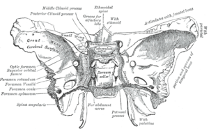
Sphenoid bone, upper surface.
|
|

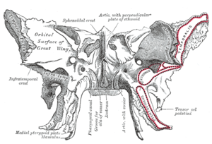
Sphenoid bone, anterior and inferior surfaces.
|
|
| Details | |
| Latin | processus pterygoideus ossis sphenoidalis |
| Identifiers | |
| Dorlands /Elsevier |
p_34/12667598 |
| TA | Lua error in Module:Wikidata at line 744: attempt to index field 'wikibase' (a nil value). |
| TH | {{#property:P1694}} |
| TE | {{#property:P1693}} |
| FMA | {{#property:P1402}} |
| Anatomical terms of bone
[[[d:Lua error in Module:Wikidata at line 863: attempt to index field 'wikibase' (a nil value).|edit on Wikidata]]]
|
|
The pterygoid processes of the sphenoid, one on either side, descend perpendicularly from the regions where the body and the greater wings of the sphenoid bone unite.
Each process consists of a medial pterygoid plate and a lateral pterygoid plate, which serve as the origins of the medial and lateral pterygoid muscles, respectively. The medial pterygoid, along with the masseter allows the jaw to move in a vertical direction as it contracts and relaxes. The lateral pterygoid allows the jaw to move in a horizontal direction during mastication. Fracture of either plate are used in clinical medicine to distinguish the Le Fort fracture classification for high impact injuries to the sphenoid and maxillary bones.
The superior portion of the pterygoid processes are fused anteriorly; a vertical groove, the pterygopalatine fossa, descends on the front of the line of fusion. The plates are separated below by an angular cleft, the pterygoid notch, the margins of which are rough for articulation with the pyramidal process of the palatine bone.
The two plates diverge behind and enclose between them a V-shaped fossa, the pterygoid fossa, which contains the medial pterygoid muscle and the tensor veli palatini.
Above this fossa is a small, oval, shallow depression, the scaphoid fossa, which gives origin to the tensor veli palatini.
The anterior surface of the pterygoid process is broad and triangular near its root, where it forms the posterior wall of the pterygopalatine fossa and presents the anterior orifice of the pterygoid canal.
In many mammals it remains as a separate bone called the pterygoid bone.
Its name is Greek for "resembling a fin or wing", from its shape.
Additional images
-
Gray187.png
Base of skull. Inferior surface.
References
This article incorporates text in the public domain from the 20th edition of Gray's Anatomy (1918)
External links
- Anatomy diagram: 34257.000-1 at Roche Lexicon - illustrated navigator, Elsevier

