Papers by Devendra P . S . Negi
Colloids and Surfaces A: Physicochemical and Engineering Aspects, 2018
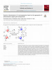
The purpose of the present work was to design a colorimetric method for the determination of 6-me... more The purpose of the present work was to design a colorimetric method for the determination of 6-mercaptopurine (6-MP). 6-MP is a useful drug for the treatment of leukemia. Gold nanoparticles (NPs) are known to undergo a change in colour upon aggregation. Hence, in the present work phenylalanine-capped gold NPs were used as a colorimetric probe for the determination of 6-MP. Transmission electron microscopy (TEM) measurements revealed that the phenylalanine-capped gold NPs ranged from 6 to 9 nm in size. The UV-vis spectrophotometric measurements showed that the surface plasmon resonance (SPR) band of the metallic NPs was positioned at 516 nm. The effect of 6-MP addition on the SPR band of the gold NPs was investigated at pH 8 and 5. At pH 8, only a slight change in the position and intensity of the SPR band was observed. However, at pH 5, the SPR band of the gold NPs was broadened as well as red shifted in the presence of 6-MP. The colour of the gold NPs changed from red to dark purple. The ratio of the absorbance of the gold NPs at 600 and 516 nm versus the concentration of 6-MP was found to be linear in the range 0.2-1.8 μM. The limit of detection (LOD) of the colorimetric method for the determination of 6-MP was calculated to be 0.647 μM.

Spectrochimica Acta A, 2020
Herein, colloidal CdS QDs have been synthesized by using cysteine as a stabilizing agent. The int... more Herein, colloidal CdS QDs have been synthesized by using cysteine as a stabilizing agent. The interaction between the CdS QDs and selenious acid was monitored by using UV-visible, fluorescence and Fourier transform infrared (FTIR) spectroscopy. The onset of absorption of the CdS QDs (430 nm) was progressively red-shifted upon increase in the concentration of selenious acid at pH 10. It indicated enlargement of the particle size which was confirmed by the dynamic light scattering (DLS) measurements. Interestingly, the addition of 100 μM selenious acid at pH 6 resulted in a 6-fold enhancement of the red emission (λ max = 617 nm) of the CdS QDs. The particle size enlargement of CdS was due to an electrostatic interaction between selenious acid and QD stabilizer cysteine. The 6-fold fluorescence enhancement was of the CdS QDs was explained on the basis of hydrogen-bonding interaction between selenious acid and cysteine. The fluorescence-based method was applied for the sensing of selenious acid at pH 6.

Materials Research Express, 2014
In this paper, histidine-stabilized ZnS nanospheres have been used as a fluorescent probe for the... more In this paper, histidine-stabilized ZnS nanospheres have been used as a fluorescent probe for the selective detection of 1,2,4 –trihydroxybenzene (1,2,4 –THB). 1,2,4 –THB quenches the fluorescence of ZnS nanospheres in the 0.2–1.0 μM concentration range. Two other isomers of the analyte (namely, 1,2,3 –THB and 1,3,5 –THB) do not quench the fluorescence of the ZnS nanospheres in the above mentioned concentration range. A charge transfer complex is formed between 1,2,4 –THB and the histidine molecules surrounding the ZnS nanospheres. Due to its electron-rich character, 1,2,4 –THB acts as an electron donor while histidine acts as an electron acceptor. The limit of detection (LOD) of 1,2,4 –THB using this method is 0.28 μM. This fluorescence-based method has the potential to be used for the detection of 1,2,4 –THB in biological samples since the experiments have been carried out in an aqueous medium at pH 7.2.

Nanotechnology, Jan 17, 2011
We have used fluorescent ZnS nanoparticles as a probe for the determination of adenine. A typical... more We have used fluorescent ZnS nanoparticles as a probe for the determination of adenine. A typical 2 × 10(-7) M concentration of adenine quenches 39.3% of the ZnS fluorescence. The decrease in ZnS fluorescence as a function of adenine concentration was found to be linear in the concentration range 5 × 10(-9)-2 × 10(-7) M. The limit of detection (LOD) of adenine by this method is 3 nM. Among the DNA bases, only adenine quenched the fluorescence of ZnS nanoparticles in the submicromolar concentration range, thus adding selectivity to the method. The amino group of adenine was important in determining the quenching efficiency. Steady-state fluorescence experiments suggest that one molecule of adenine is sufficient to quench the emission arising from a cluster of ZnS consisting of about 20 molecules. Time-resolved fluorescence measurements indicate that the adenine molecules block the sites on the surface of ZnS responsible for emission with the longest lifetime component. This method ma...
Chemical Physics Letters, 2010
Cadmium sulfide particles have been synthesized in the aqueous medium using the amino acid histid... more Cadmium sulfide particles have been synthesized in the aqueous medium using the amino acid histidine as a stabilizing agent. These particles demonstrate the phenomenon of size quantization effect. The fluorescence of histidine-stabilized CdS was found to be enhanced and quenched by the addition of DNA bases adenine and guanine, respectively. The fluorescence enhancement of CdS in the presence of adenine
Colloidal silver nanoparticles (AgNPs) have been synthesized using polyvinylpyrrolidone (PVP) as ... more Colloidal silver nanoparticles (AgNPs) have been synthesized using polyvinylpyrrolidone (PVP) as a capping agent. The particles were characterized using UV-visible absorption spectroscopy and transmission electron microscopy (TEM). The particles were 4-12 nm in diameter. The surface plasmon resonance (SPR) band of the AgNPs at 400 nm was quenched in the presence of 1 μM Pb2+ ions at pH 9.6. Additionally, a new absorption band was formed around 600 nm. The colour of the colloidal solution was found to change from yellow to green. The addition of metal ions such as Al3+, Ca2+, Cd2+, Co2+, Cu2+, Hg2+, Mg2+, Ni2+, Sm3+ and Zn2+ did not alter the SPR band of the AgNPs significantly. The limit of detection (LOD) of Pb2+ ions by the colorimetric method was found to be 14.4 nM. The mechanism of the aggregation of the AgNPs in the presence of the Pb2+ ions has been discussed.

Zinc sulfide nanoparticles have been synthesized using sodium dodecylsulfate micelles as a stabil... more Zinc sulfide nanoparticles have been synthesized using sodium dodecylsulfate micelles as a stabilizing agent. The as-prepared nanoparticles are characterized by ultraviolet-visible spectroscopy, energy dispersive X-ray spectroscopy, X-ray diffraction and transmission electron microscopy. The photocatalytic activity of the ZnS nanoparticles has been evaluated by monitoring the degradation of methyl orange with a UV-visible spectrophotometer. The effect of ZnS concentration on the photocatalytic degradation of the dye has been investigated. Under optimum experimental conditions, 98% of the dye is degraded in the presence of the photocatalyst during 10 min of irradiation with ultraviolet light. The turnover frequency for methyl orange degradation per mg of ZnS is calculated to be 6×10 molecules s. The effect of SDS concentration on the photocatalytic degradation of the dye has also been investigated. The results suggest that SDS micelles do not catalyze the degradation of the dye. The ...

Journal of Chemical Sciences, 2002
An intramolecular charge transfer (ICT) molecule, p-N,N-dimethylaminobenzoic acid (DMABA) has bee... more An intramolecular charge transfer (ICT) molecule, p-N,N-dimethylaminobenzoic acid (DMABA) has been studied in zeolite and colloidal media. The ratio of ICT to normal emission (ICT/LE) is greatly enhanced in zeolites compared to that in polar solvents. The ICT emission of DMABA was quenched by increasing the concentration of TiO 2 colloids, while the normal emission was slightly enhanced. Upon illumination of the heteropoly acid (HPA) incorporated TiO 2 colloids, interfacial electron transfer takes place from the conduction band of TiO 2 to the incorporated HPA which is also excited to catalyze the photoreduction of Methyl Orange. It is found that the interfacial electron transfer mechanism of HPA/TiO 2 is quite analogous to the Z-scheme mechanism for plant photosynthetic systems. In DMABA-adsorbed TiO 2 /Y-zeolite the ICT/LE ratio of DMABA is quite small implying that electron transfer takes place from DMABA to the conduction band of TiO 2. This results in drastic enhancement in the photocatalytic activity of DMABAadsorbed TiO 2 /Y-zeolite compared to free TiO 2 /Y-zeolite.

In the present work we have investigated the oxidation of L-3,4-dihydroxyphenylalanine (L-Dopa) u... more In the present work we have investigated the oxidation of L-3,4-dihydroxyphenylalanine (L-Dopa) using colloidal cadmium sulfide nanoparticles (CdS NPs) as a photocatalyst. The CdS NPs were synthesized and characterized using UV-visible absorption spectroscopy, transmission electron microscopy (TEM) and powder x-ray diffraction (XRD). It was observed that the CdS NPs oxidized L-Dopa within 30 min of irradiation using a 200 W Hg(Xe) arc lamp. The oxidation product was identified using high performance liquid chromatography (HPLC). The HPLC analyses were carried out on a reverse-phase C18 column under isocratic conditions using methanol/water (10/90) as the mobile phase. The retention time of the product matched with that of dopachrome. The mechanistic studies indicated the participation of hydroxyl radicals (• OH) in the photocatalytic oxidation of L-Dopa. The kinetic analysis was carried out using the Michaelis-Menten equation. The results demonstrated that the as-prepared CdS NPs mimicked the activity of the catechol oxidase enzyme. To the best of our knowledge, this is the first report on the oxidation of L-Dopa using semiconductor NPs.

The purpose of the present research was to design a method for the colorimetric determination of ... more The purpose of the present research was to design a method for the colorimetric determination of cysteamine. We have employed cysteine-stabilized silver nanoparticles (AgNPs) as a probe. The addition of cysteamine resulted in the quenching of the 400 nm surface plasmon resonance (SPR) band of the AgNPs. It was accompanied by the appearance of a new absorption band at 560 nm. The colour of the colloidal AgNPs changed from yellow to dark brown within a few seconds. The change in colour of the AgNPs was due to their aggregation induced by the addition of cysteamine. Significantly, other biomolecules such as arginine, asparagine, aspartic acid, cysteine, glutamic acid, glutathione, glycine, methionine and 6-mercaptopurine did not cause any change in the colour of the AgNPs. The limit of detection (LOD) of the method was 0.37 μM. The mechanism of the aggregation of the AgNPs induced by cysteamine has also been described. The method has been applied for the detection of cysteamine in human blood serum.

Journal of Molecular Liquids, 2019
The purpose of the current research was to study the interaction between the DNA nucleobases (hyp... more The purpose of the current research was to study the interaction between the DNA nucleobases (hypoxanthine and guanine) and the ZnS nanoparticles (NPs). A good understanding of such interactions could be beneficial to researchers working in the field of bionanotechnology. The ZnS NPs were synthesized by a reported method and characterized by powder X-ray diffraction (XRD) and transmission electron microscopy (TEM). The interaction between the DNA nucleobases and the ZnS NPs was investigated by using UV-visible, fluorescence and in-frared spectroscopy. The experiments were performed at pH 6.0 and 10.0 to investigate the role of the molecular charge on the interaction process. The UV-visible absorption studies revealed that the absorption bands of the DNA nucleobases were red shifted in the presence of the semiconductor NPs. The red shift was observed to be greater at pH 6.0. The steady-state fluorescence studies showed that the nucleobases quenched the fluorescence emission of ZnS. The quenching was found to depend on the pH of the solution. It was observed that hypoxanthine was more efficient in quenching the fluorescence of the ZnS NPs compared to guanine. The infra-red spectroscopic measurements indicated that both hypoxanthine and guanine bonded to the semiconductor surface through their nitrogen atoms.
Journal of Chemical Research (Synopses), 1998
Binding of 3-methylindole (3-MI) to the surface of colloidal CdS particles modifies their lumines... more Binding of 3-methylindole (3-MI) to the surface of colloidal CdS particles modifies their luminescence behaviour so that the trapped electron and hole generated upon photoirradiation are scavenged by adsorbed O 2 and 3-MI to yield 2-acetylformanilide and 2-aminoacetophenone.
Journal of Colloid and Interface Science, 2001
The photophysics of Cd(OH) 2-coated Q-CdS in the presence of guanine and related compounds has be... more The photophysics of Cd(OH) 2-coated Q-CdS in the presence of guanine and related compounds has been examined. Guanine and adenine quench the bandgap emission and reduce the emission lifetime of these particles. Approximately 50% of the bandgap flu-orescence is quenched by a low [guanine] (2×10 −5 mol dm −3). Quenching takes place with a bimolecular rate constant of 2× 10 12 dm 3 mol −1 s −1. The presence of these additives did not affect the red emission appreciably. The nature of the interaction between Cd(OH) 2 layer of Q-CdS and the additive has been analyzed by fluorescence and absorption spectroscopy. Interception of the hole by guanine and adenine occurs through their adsorption and hydrogen-bonding interaction between the-OH of Cd(OH) 2 and certain functional groups of the additive. Cd(OH) 2-coated Q-CdS sensitizes the photodecomposition of guanine efficiently in the presence of oxygen under visible light irradiation. Shallowly trapped and direct holes are suggested to participate in the oxidation. The reactivity of the hole is governed by the redox potential of the solute. C 2001 Academic Press

Journal of Photochemistry and Photobiology A, 2000
Coating of Cd(OH) 2 on Q-CdS particles enhances their photostability, luminescing efficiency and ... more Coating of Cd(OH) 2 on Q-CdS particles enhances their photostability, luminescing efficiency and emission lifetime. The presence of tryptophan quenches the bandgap emission of CdS and reduces its emission lifetime. For a typical 2×10 −4 mol dm −3 of tryptophan the average emission lifetime of CdS is reduced from 22.6 to 8.5 ns. For this process a quenching rate constant of about 4×10 11 dm 3 mol −1 s −1 has been evaluated. The red emission is not influenced appreciably. Cd(OH) 2-coated Q-CdS sensitised photooxidation of tryptophan has been examined in the presence of oxygen by irradiating with visible light. The photogenerated hole on the particle is intercepted by the bulk substrate (φ −tryp =0.22) to produce 5-hydroxytryptophan (φ 5-OHtryp =0.08) as one of the main products. Shallowly trapped hole has been assigned to participate in the oxidation via hydrogen bonding interaction involving the surface of the particle and the substrate. Emission experiments indicate a difference in nature of reactive hole photogenerated on stoichiometric and Cd(OH) 2-coated Q-CdS particles. A mechanism of this reaction has been proposed.
Nanotechnology, 2008
We present a novel method for the selective detection of cysteine, a sulfur-containing amino acid... more We present a novel method for the selective detection of cysteine, a sulfur-containing amino acid, which plays a crucial role in many important biological functions such as protein folding. Surface-modified colloidal CdS nanoparticles have been used as a fluorescent probe to selectively detect cysteine in the presence of other amino acids in the micromolar concentration range. Cysteine quenches the emission of CdS in the 0.5-10 μM concentration range, whereas the other amino acids do not affect its emission. Among the other amino acids, histidine is most efficient in quenching the emission of the CdS nanoparticles. The sulfur atom of cysteine plays a crucial role in the quenching process in the 0.5-10 μM concentration range. Cysteine is believed to quench the emission of the CdS nanoparticles by binding to their surface via its negatively charged sulfur atom. This method can potentially be applied for its detection in biological samples.

Materials Research Express, 2016
Gold nanoparticles (AuNPs) have been synthesized using ethylenediaminetetraacetic acid (EDTA) as ... more Gold nanoparticles (AuNPs) have been synthesized using ethylenediaminetetraacetic acid (EDTA) as a capping agent. Transmission electron microscopy measurements showed the particles to have an average size of ∼40 nm. The absorption spectrum of the AuNPs displayed a sharp absorption band at 523 nm. The red colloidal solution of the AuNPs was found to immediately change its colour to blue upon addition of 20 μm Al 3+ ions. The addition of the same concentration of Cr 3+ ions resulted in the same colour change but only after ∼5 min. No colour change was observed on addition of the same concentrations of Fe 3+ , Cu 2+ , Pb 2+ , Cd 2+ , Zn 2+ , Co 2+ , Ni 2+ , Hg 2+ and Mn 2+ ions. The colour change was attributed to the aggregation of the AuNPs due to complexation of the Al 3+ ions by EDTA on the AuNPs. This was supported by dynamic light scattering data. A plot of the ratio of the extinctions at 700 and 523 nm versus the Al 3+ ion concentration was linear in the 2-18 μm range and the limit of detection was 3.6 μm.

Colloids and Surfaces A, 2018
The purpose of the present work was to design a colorimetric method for the determination of 6-me... more The purpose of the present work was to design a colorimetric method for the determination of 6-mercaptopurine (6-MP). 6-MP is a useful drug for the treatment of leukemia. Gold nanoparticles (NPs) are known to undergo a change in colour upon aggregation. Hence, in the present work phenylalanine-capped gold NPs were used as a colorimetric probe for the determination of 6-MP. Transmission electron microscopy (TEM) measurements revealed that the phenylalanine-capped gold NPs ranged from 6 to 9 nm in size. The UV-vis spectrophotometric measurements showed that the surface plasmon resonance (SPR) band of the metallic NPs was positioned at 516 nm. The effect of 6-MP addition on the SPR band of the gold NPs was investigated at pH 8 and 5. At pH 8, only a slight change in the position and intensity of the SPR band was observed. However, at pH 5, the SPR band of the gold NPs was broadened as well as red shifted in the presence of 6-MP. The colour of the gold NPs changed from red to dark purple. The ratio of the absorbance of the gold NPs at 600 and 516 nm versus the concentration of 6-MP was found to be linear in the range 0.2-1.8 μM. The limit of detection (LOD) of the colorimetric method for the determination of 6-MP was calculated to be 0.647 μM.

Spectrochimica Acta part A, 2018
The purpose of the present work was to develop a method for the sensing of thioacetamide by using... more The purpose of the present work was to develop a method for the sensing of thioacetamide by using spectroscop-ic techniques. Thioacetamide is a carcinogen and it is important to detect its presence in foodstuffs. Semiconductor quantum dots are frequently employed as sensing probes since their absorption and fluorescence properties are highly sensitive to the interaction with substrates present in the solution. In the present work, the interaction between thioacetamide and ZnO quantum dots has been investigated by using UV-visible, fluorescence and in-frared spectroscopy. Besides, dynamic light scattering (DLS) has also been utilized for the interaction studies. UV-visible absorption studies indicated the bonding of the lone pair of sulphur atom of thioacetamide with the surface of the semiconductor. The fluorescence band of the ZnO quantum dots was found to be quenched in the presence of micromolar concentrations of thioacetamide. The quenching was found to follow the Stern-Volmer relationship. The Stern-Volmer constant was evaluated to be 1.20 × 10 5 M −1. Infrared spectroscopic measurements indicated the participation of the\ \NH 2 group and the sulphur atom of thioacetamide in bonding with the surface of the ZnO quantum dots. DLS measurements indicated that the surface charge of the semiconductor was shielded by the thioacetamide molecules.


Uploads
Papers by Devendra P . S . Negi