Mri study guide
Discover Pinterest’s best ideas and inspiration for Mri study guide. Get inspired and try out new things.
61 people searched this
·
Last updated 2mo

MRI temporalis muscle anatomy

Radiological images


MRI Registry Test Preparation


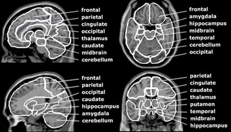
This MRI brain cross sectional anatomy tool is absolutely free to use. This section of the website will explain large and minute details of axial brain cross sectional anatomy.

CASE 1: Discussion

Learning all things #radiologicalanatomy? Master your knowledge and ability to identify structures on an #mri with our free diagrams and #anatomyquizzes. Learn more. #learnanatomy #mriscan #radiology #medicalimaging

This MRI brain cross sectional anatomy tool is absolutely free to use. This section of the website will explain large and minute details of coronal brain cross sectional anatomy.

Learning Medical Imaging, Cardiac CT to Contrast guides, Unique modules, Quiz of the month, Imaging pearls, Journal Club, Medical Illustrations, CME Courses|CTisus

This MRI wrist cross sectional anatomy tool is absolutely free to use. This section of the website will explain large and minute details of wrist coronal cross sectional anatomy.


AXIAL LUMBAR SPINE MRI ANATOMY 👨🏽💻Here we have an axial slice of the lumbar spine with T1 weighting - notice how the fluid in the…


Functional MRI studies have helped to elucidate underlying mechanisms in complex neurological and neuropsychiatric disorders. Disease processes often involve complex large-scale network interactions, extending beyond the presumed main disease focus. ...