Pretibial myxedema
| Pretibial myxedema | |
|---|---|

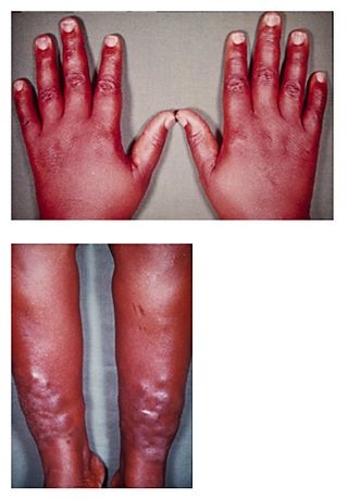
Hands showing related condition thyroid acropachy and Shins of someone with pretibial myxedema
|
|
| Classification and external resources | |
| Specialty | Lua error in Module:Wikidata at line 446: attempt to index field 'wikibase' (a nil value). |
| ICD-9-CM | 242.9 |
| DiseasesDB | 25147 |
| eMedicine | derm/347 |
| Patient UK | Pretibial myxedema |
Pretibial myxedema (myxoedema (UK), also known as Graves Dermopathy, thyroid dermopathy,[1] Jadassohn-Dösseker disease or Myxoedema tuberosum) is an infiltrative dermopathy, resulting as a rare complication of Graves' disease,[2] with an incidence rate of about 1-5% in patients.
Presentation
It usually presents itself as a waxy, discolored induration of the skin—classically described as having a so-called peau d'orange (orange peel) appearance—on the anterior aspect of the lower legs, spreading to the dorsum of the feet, or as a non-localised, non-pitting edema of the skin in the same areas.[3] In advanced cases, this may extend to the upper trunk (torso), upper extremities, face, neck, back, chest and ears.
The lesions are known to resolve very slowly. Application of petroleum jelly on the affected area could relieve the burning sensation and the itching. It occasionally occurs in non-thyrotoxic Graves' disease, Hashimoto's thyroiditis, and stasis dermatitis. The serum contains circulating factors which stimulate fibroblasts to increase synthesis of glycosaminoglycans.
Risk Factors
There are suggestions in the medical literature that treatment with radioactive iodine for Graves' hyperthyroidism may be a trigger for pretibial myxedema[4] which would be consistent with radioiodine ablation causing or aggravating ophthalmopathy, a condition which commonly occurs with pretibial myxedema and is believed to have common underlying features.[5]
Other known triggers for ophthalmopathy include thyroid hormone imbalance, and tobacco smoking, but there has been little research attempting to confirm these are also risk factors for pretibial myxedema.
Diagnosis
A biopsy of the affected skin reveals mucin in the mid- to lower- dermis. There is no increase in fibroblasts. Over time, secondary hyperkeratosis may occur, which may become verruciform. Many of these patients may also have co-existing stasis dermatitis. Elastic stains will reveal a reduction in elastic tissue.
References
- ↑ Lua error in package.lua at line 80: module 'strict' not found.
- ↑ Lua error in package.lua at line 80: module 'strict' not found.
- ↑ Lua error in package.lua at line 80: module 'strict' not found.
- ↑ Lua error in package.lua at line 80: module 'strict' not found.
- ↑ Lua error in package.lua at line 80: module 'strict' not found.