

White ramus communicans
| White ramus communicans | |
|---|---|
 Scheme showing structure of a typical spinal nerve. 1. Somatic efferent. 2. Somatic afferent. 3,4,5. Sympathetic efferent. 6,7. Sympathetic afferent. | |
 Diagram of the course and branches of a typical intercostal nerve. (Rami communicantes labeled at center.) | |
| Details | |
| Identifiers | |
| Latin | ramus communicans albus nervi spinalis |
| TA98 | A14.3.01.007 |
| TA2 | 6150 |
| FMA | 5875 |
| Anatomical terminology | |
The thoracic, and the first and second lumbar nerves each contribute a branch, white ramus communicans to the adjoining sympathetic ganglion.
They contain both myelinated and unmyelinated preganglionic sympathetic fibers, (GVE and GVA). The white ramus appears white because there are more myelinated than unmyelinated fibres unlike the gray rami.
Unlike the gray rami, white rami communicantes do not extend below L2 or above T1.[1]
Additional Info
-
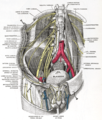 Deep and superficial dissection of the lumbar plexus.
 -
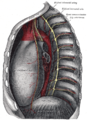 Thoracic portion of the sympathetic trunk.


The white rami communicantes (plural of ramus communicans, ramus means branch) is the preganglionic sympathetic outflow from the spinal cord.
The cell bodies for the preganglionic sympathetic myelinated fibers in the white rami communicantes lie in the ipsilateral (same sided) intermediolateral cell column in the spinal cord which extends from T1-L2. These rami also contain general visceral afferent fibers (sensory from the organs) whose primary cell bodies reside dorsal root ganglia (which then synapse in the anterior horn). The preganglionic sympathetic fibers will enter into the sympathetic trunk and either synapse at the ganglion on the same level, or travel up or down the sympathetic trunk to arrive at the correct spinal level for their action. Once they synapse in the sympathetic ganglion in the sympathetic trunk, they exit the trunk as gray rami to join the spinal nerve and innervate the appropriate structure. Even though the sympathetic trunk extends below L2, there are no more white rami communicantes below L2 because the intermediolateral cell column ends before this. The fibers of the sympathetic trunk above and below T1-L2 originate from white rami communicantes within T1-L2. Above and below T1-L2 there are only gray rami.
See also
References
- ^ "Dissector Answers - Kidney & Retroperitoneum". Retrieved 2007-11-13.
External links
- Template:EMedicineDictionary
- Atlas image: n3a6p1 at the University of Michigan Health System - "Autonomic Connections of the Spinal Cord"
- Overview and diagram
![]() This article incorporates text in the public domain from page 920 of the 20th edition of Gray's Anatomy (1918)
This article incorporates text in the public domain from page 920 of the 20th edition of Gray's Anatomy (1918)
Anatomy of the autonomic nervous system | |||||
|---|---|---|---|---|---|
| Head |
| ||||
| Neck |
| ||||
| Thorax |
| ||||
| Abdomen |
| ||||
| Pelvis |
| ||||
This neuroscience article is a stub. You can help Wikipedia by expanding it. |