Thesis Chapters by Dr. Md. Aktaruzzaman
Papers by Dr. Md. Aktaruzzaman

Aster spathulifolius Maxim. is a perennial herb in the family Asteraceae, which is narrowly distr... more Aster spathulifolius Maxim. is a perennial herb in the family Asteraceae, which is narrowly distributed and endemic in coastal regions of Korea and Japan (Nguyen et al. 2013). This plant has long been grown in gardens for ornamental purposes in Korea and has recently been valued as a medicinal herb (Won et al. 2013). In August 2016, dozens of A. spathulifolius plants showing symptoms of powdery mildew were observed in the natural habitat of Ulleung island (37°29'30"N; 130°54'39"E), Korea, with a disease incidence of ca. 10%. The powdery mildew colonies were circular to irregular, forming white patches on both sides of the leaves. The voucher specimens were deposited in the Korea University herbarium (KUS-F23869 and F29401). Hyphae were flexuous to straight, branched, septate, and 4 to 7 μm wide. Hyphal appressoria were nipple-shaped or nearly absent. Conidiophores were straight, 135 to 250 × 10 to 12 μm and formed apically 2 to 4 immature conidia in chains showing crenate outline. Foot-cells of conidiophores were cylindrical and 35 to 65 μm long. Conidia were ellipsoid-ovoid to barrel-shaped, 27 to 40 × 18 to 23 μm (length/width ratio of 1.5 to 2.2) and containing distinct fibrosin bodies. Teleomorph stage of the fungus was not observed. All morphological features described above are typical of the powdery mildew Fibroidium anamorph of the genus Podosphaera. Aiming to confirm the fungus identity, internal transcribed spacer (ITS) region of KUS-F29401 was amplified with the primers ITS5 and P3 as described by Takamatsu et al. (2009) and sequenced directly. A BLAST search of the Korean isolate had a high similarity (>99%) with those of P. astericola on asteraceous hosts (AB040353, AB040335, AB040341, and KU147187) and the next closest taxon was P. carpesiicola (AB040350). The obtained 775 bp sequence
In this study, we identified the causative agent of post-harvest gray mold on broccoli that was s... more In this study, we identified the causative agent of post-harvest gray mold on broccoli that was stored on a farmers' cooperative in Pyeongchang, Gangwon Province, South Korea, in September 2016. The incidence of gray mold on broccoli was 10-30% after 3-5 weeks of storage at 3 o C. Symptoms included brownish curd and gray-to-dark mycelia with abundant conidia on the infected broccoli curds. The fungus was isolated from infected fruit and cultured on potato dextrose agar. To identify the fungus, we examined the morphological characteristics and sequenced the rDNA of the fungus and confirmed its pathogenicity according to Koch's postulates. The results of the morphological examination, pathogenicity test, and sequencing of the 5.8S rDNA of the internal transcribed spacer regions (ITS1 and ITS4) and three nuclear protein-coding genes, G3P-DH, HSP60, and RPB2, revealed that the causal agent of the post-harvest gray mold on broccoli was Botrytis cinerea. To our knowledge, this is the first report of post-harvest gray mold on broccoli in Korea.

PDF Print | PDF with Links Strawflower (Xerochrysum bracteatum) is a flowering plant of the Aster... more PDF Print | PDF with Links Strawflower (Xerochrysum bracteatum) is a flowering plant of the Asteraceae. In July 2016, approximately 10% of inflorescences were observed displaying gray mold symptoms in a private garden in Gangneung, Korea. Initial infection started from the base of inflorescence and gradually moved to the flower spike where it finally collapsed. Lesions expanded rapidly under cool, humid conditions. Diseased tissue was excised and surface sterilized by immersing in 1% sodium hypochlorite (NaOCl) for 1 min, rinsed three times with sterilized distilled water, placed on potato dextrose agar (PDA, Difco) plates, and incubated at 20 ± 2°C for 7 days. The fungus produced gray to grayish brown colonies that sporulated abundantly. The conidia (n = 50) were one-celled, ellipsoid or ovoid, dark brown, and 5.1 to 7.2 × 5.3 to 5.6 μm in vivo, and 5.1 to 10.4 × 5.1 to 7.8 μm in vitro. Conidiophores (n = 20) arose solitary or in groups, straight or flexuous, septate, with an inflated basal cell brown to light brown, and measured 110 to 406 × 10 to 27 μm. After 21 days, the fungus formed several black sclerotia (n = 20) ranging from 1.1 to 4.3 × 1.2 to 3.5 mm near the edge of the Petri dish. A representative isolate (SFGM002) was deposited
In May 2016, an occurrence of grey mould rots was observed on harvested green bean pods after 3–5... more In May 2016, an occurrence of grey mould rots was observed on harvested green bean pods after 3–5 days of storage in refrigerator in Gangneung, Gangwon Province, South Korea. The symptoms observed were water-soaked grey le-sions with white to greyish mycelium on infected pods. The fungus was isolated from infected pods and cultured on potato dextrose agar (PDA). For identification of the fungus, we examined morphology, sequenced a portion of the rDNA and three nuclear protein coding genes and confirmed its pathoge-nicity according to Koch's postulates. The results of morphological examinations, pathogenicity tests and DNA revealed that the causal agent was Botrytis cinerea. This confirms post-harvest grey mould rot of green bean in Korea.
In October 2016, we collected samples of
post-harvest anthracnose of papaya from Gangneung,
Gangw... more In October 2016, we collected samples of
post-harvest anthracnose of papaya from Gangneung,
Gangwon Province, Korea. Infected fruits first showed
tiny light brown spots on their surface followed by
development of sunken water-soaked lesions with black
acervuli. Fungal pathogen was isolated from the infected
fruits and cultured on potato dextrose agar (PDA) and
synthetic nutrient agar (SNA) medium. For identification
of the fungus, morphological assessment and sequencing
analysis of 5.8S ribosomal DNA (ITS-5.8S
rDNA) and three protein-coding genes: actin (ACT),
chitin synthase I (CHS-1), and glyceraldehyde-3-
phosphate dehydrogenase (G3PDH) were performed.
Pathogenicity of the fungal isolate was confirmed according
to Koch’s postulates. The results of morphological
examination, pathogenicity tests, and the ITS-5.8S
rDNA, ACT, CHS-1, and G3PDH sequences confirmed
Colletotrichum truncatum as the causal agent. This
study provides news information about C. truncatum
isolated from post-harvest papaya in Korea.
In this study, we identified the causative
agent of postharvest gray-mold rot in sweet persimmon
... more In this study, we identified the causative
agent of postharvest gray-mold rot in sweet persimmon
fruit that were collected from Gangneung,
Gangwon Province, Korea in October 2016. Symptoms
included extensive growth of mycelia on post
harvested fruit. The fungus was isolated from infected
fruit and cultured on potato dextrose agar
(PDA). For identification of the fungus, we examined
morphology characteristics and rDNA sequencing
analysis of the fungus and confirmed its
pathogenicity according to Koch’s postulates. The
results of morphological examinations, pathogenicity
tests, 5.8S rDNA sequences of the internal transcribed
spacer regions (ITS1 and ITS4) and the five
nuclear protein-coding genes G3PDH, HSP60,
RPB2, MS547 and TUB revealed that the causal
agent of postharvest gray-mold rot on sweet persimmon
fruit in Korea was Botrytis cinerea.
In May 2016, we observed gray mold rot symptoms on harvested and refrigerated cherry fruit in Gan... more In May 2016, we observed gray mold rot symptoms on harvested and refrigerated cherry fruit in Gangneung, Gangwon Province, Korea. The symptoms included extensive growth of mycelia with gray conidia on infected fruit. We isolated the pathogen from infected fruit and cultured the fungus on potato dextrose agar. For identification of the fungus, we examined its morphology and rDNA sequence and confirmed its pathogenicity according to Koch's postulates. The results of morphological examinations, pathogenicity tests, and rDNA sequence analysis of internal transcribed spacer regions 1 and 4, and three nuclear protein-coding genes, glycer-aldehyde-3-phosphate dehydrogenase gene, heat shock protein 60 gene, and DNA-dependent RNA polymerase subunit II gene revealed that the causal agent was Botrytis cinerea. To our knowledge, this is the first report of postharvest gray mold rot of sweet cherry in Korea.
In October 2014, an occurrence of gray mold was observed on young fruits of sponge gourd
(Luffa c... more In October 2014, an occurrence of gray mold was observed on young fruits of sponge gourd
(Luffa cylindrica) in Sachunmun, Gangneung, South Korea. Symptoms included abundant mycelia
growth with gray conidia on young fruits and finally rotting the fruits. The fungus was isolated from
symptomatic fruits and its pathogenicity was confirmed. Based on the morphological features and
sequence analysis of ITS-5.8S rDNA, G3PDH, HSP60, and RPB2 genes, the pathogen was identified as
Botrytis cinerea Pers. This is the first report of gray mold caused by B. cinerea on L. cylindrica in Korea.
Crepe myrtle (Lagerstroemia indica, Lythraceae) is a flowering shrub widely planted in warmer cli... more Crepe myrtle (Lagerstroemia indica, Lythraceae) is a flowering shrub widely planted in warmer climate zones as landscape plant, generally producing diverse colored flowers in summer and early fall (Qin and Graham 2007). In Korea, gray mold disease was first observed during the rainy season in 2013 in Gangneung, Korea. In 2014 the disease was not aggressive, but in July 2015 approximately 5 to 10% of inflorescences displayed symptoms of gray mold that reduced the aesthetic value of flowers. Watersoaked, brown or gray spots followed by abundant mycelia with conidia appeared on the affected inflorescences. Diseased tissue was surfacesterilized by immersing in 0.1% sodium hypochlorite (NaOCl) for 1 min, rinsed three times with sterile water, placed on potato dextrose agar (PDA, Difco), and incubated at 20 ± 2°C. The fungus formed black sclerotia ranging from 1.1 to 4.4 × 1.0 to 3.2 mm (n = 20) near the edges of culture dishes. Conidia (n = 50) were ellipsoidal or ovoid, 5.6 to 8.1 × 5.7 to 9.6 µm on naturally infected inflorescences and 5.8 to 10.6 × 5.3 to 7.5 µm on PDA. Morphological characteristics were
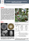
In 2013, gray mold symptoms were frequently observed on red raspberry (Rubus idaeus L.) in Gangnu... more In 2013, gray mold symptoms were frequently observed on red raspberry (Rubus idaeus L.) in Gangnueng, Gangwon Province in Korea. The typical symptoms were dark brown lesions covered with green-gray spore masses appeared on twigs, blossoms, leaves and fruit. Lesions expanded rapidly under cool, humid condition. A fungal isolate was obtained from symptomatic on infected plant parts. Conidia were pale brown, one-celled, mostly ellipsoid or ovoid in shape and were 5.9~10.8 × 4.9~7.3 ㎛ in size. Black, irregular sclerotia formed at random on potato dextrose agar (PDA) over time. The optimal temperature for mycelial growth and sclerotia formation of the fungal isolates was 20℃. The causal fungus of gray mold isolated from the diseased plant was identified as Botrytis cinerea based on mycological characteristics and ITS rDNA sequences. This is the first report on gray mold of red raspberry caused by B. cinerea in Korea. ABSTRACT RESULTS
In August 2015, we collected samples of gray mold from sweet basil growing in Sachunmeon, Gangneu... more In August 2015, we collected samples of gray mold from sweet basil growing in Sachunmeon, Gangneung,
Gangwon Province, Korea. Symptoms included extensive growth of mycelia with gray conidia on young leaves, stems, and
blossoms. The pathogen was isolated from infected leaves and blossoms and the fungus was cultured on potato dextrose agar. For
identification of the fungus, morphology and rDNA sequencing analysis of the fungus were performed, which confirmed its
pathogenicity according to Koch’s postulates. The results of morphological examinations, pathogenicity tests, and the rDNA
sequences of the internal transcribed spacer regions (ITS1 and ITS4) and the three nuclear protein-coding genes G3PDH, HSP60,
and RPB2 showed that the causal agent was Botrytis cinerea. This is the first report of gray mold caused by Botrytis cinerea on
sweet basil in Korea.
In June 2013, we collected leaf spot disease samples of Datura metel from Gangneung, Gangwon Prov... more In June 2013, we collected leaf spot disease samples of Datura metel from Gangneung, Gangwon Province,
Korea. The symptoms observed were small circular to oval dark brown spots with irregular in shape
or remained circular with concentric rings. We isolated the pathogen from infected leaves and cultured the
fungus on potato dextrose agar. We examined the fungus morphologically and confirmed its pathogenicity
according to Koch’s postulates. The results of morphological examinations, pathogenicity tests, and the rDNA
sequences of the internal transcribed spacer regions (ITS1 and ITS4), glycerol-3-phosphate dehydrogenase
(G3PDH) and the RNA polymerase II second largest subunit (RPB2) gene sequence revealed that the causal
agent was Alternaria tenuissima. To the best of our knowledge, this is the first report of leaf spot of D. metel
caused by A. tenuissima in Korea as well as worldwide.

During May and June 2014, red raspberry (Rubus idaeus L., Rosaceae) plants with symptoms resembli... more During May and June 2014, red raspberry (Rubus idaeus L., Rosaceae) plants with symptoms resembling gray mold were found in several plastic greenhouses with approximately 2 to 10% disease incidence (percentage of inflorescences infected) in Gangneung, Korea. Symptoms included extensive growth of mycelia with gray conidia, mostly on blossoms and fruits, rarely on leaves and twigs. Diseased fruit tissue was excised and surface sterilized by immersion in 0.1% sodium hypochlorite (NaOCl) for 1 min, rinsed three times with sterilized distilled water, placed on potato dextrose agar (PDA, Difco) plates, and incubated at 20 ± 2°C. Conidia (n = 50) were single-celled, ellipsoidal or ovoid, 6.3 to 8.2 × 5.7 to 9.7 μm on naturally infected fruits and 5.9 to 10.8 × 4.9 to 7.3 μm on PDA. Monoconidial isolates were obtained by plating conidial suspensions on PDA and selecting single colonies. After three weeks, the fungus formed several black sclerotia ranging from 1.1 to 4.2 × 1.3 to 3.2 mm (n = 20) near the edge of the Petri dish. A representative isolate (GRGM003) was deposited in Gangneung-Wonju National University and used for further studies. Morphological characteristics were consistent with those of Botrytis cinerea Pers.: Fr. (Ellis 1971). To confirm the identity of the causal fungus, the internal transcribed spacer (ITS) region of rDNA of the isolate was amplified with universal primers ITS1/ITS4 and sequenced. BLAST analysis of the resulting 500-bp nucleotide ITS segment (GenBank, KP255842) showed 100% identity with the sequence of Botryotinia fuckeliana (teleomorph of Botrytis cinerea). For further confirmation, three nuclear protein-coding genes were sequenced: glyceraldehyde-3-phosphate dehydrogenase (G3PDH), heat-shock protein 60 (HSP60), and DNA-dependent RNA polymerase subunit II (RPB2) (Staats et al. 2005). The G3PDH, HSP60, and RPB2 sequences (KT025631, LC020016, and KT025632) of the isolate were 100% identical to those of B. cinerea (e.g., KF015583, KJ018758, and KJ018756, respectively). For the pathogenicity test, inoculum was prepared by harvesting conidia from 2-week-old cultures on PDA. A suspension (1×10⁶ conidia/mL) amended with 0.5% glucose and 0.25% K2HPO4 was sprayed onto three potted red raspberry plants with fruits. Three plants of similar features were sprayed with sterilized water, serving as controls. Plants were covered with plastic bags for 2 days after inoculation to maintain high relative humidity (90 ± 10%) and were placed in a growth chamber at 20 ± 2°C. Gray mold symptoms on fruits and leaves developed 3 to 5 days after inoculation, whereas control plants remained symptomless. The pathogenicity test was performed twice with similar results. The pathogen was successfully re-isolated from inoculated leaves, fulfilling Koch’s postulates. Red raspberry gray mold has been reported in many countries including the United States and China (Farr and Rossman 2015). To our knowledge, this is the first report of gray mold caused by B. cinerea on red raspberry in Korea. According to our observations, fruit infections occurred under high humidity of the greenhouse. We suppose that ventilation of the greenhouses may effectively reduce severity of gray mold on raspberry fruits.
Herein, we report the first occurrence of web blight of rosemary caused by Rhizoctonia solani AG-... more Herein, we report the first occurrence of web blight of rosemary caused by Rhizoctonia solani AG-1-IB in Gangneung,
Gangwon Province, Korea, in August 2014. The leaf tissues of infected rosemary plants were blighted and white mycelial growth
was seen on the stems. The fungus was isolated from diseased leaf tissue and cultured on potato dextrose agar for identification.
The young hyphae had acute angular branching near the distal septum of the multinucleate cells and mature hyphal branches
formed at an approximately 90o angle. This is morphologically identical to R. solani AG-1-IB, as per previous reports. rDNA-ITS
sequences of the fungus were homologous to those of R. solani AG-1-IB isolates in the GenBank database with a similarity
percentage of 99%, thereby confirming the identity of the causative agent of the disease. Pathogenicity of the fungus in rosemary
plants was also confirmed by Koch’s postulates.

In April 2014, hundreds of ready-to-market cineraria [Senecio cruentus (Masson ex L’Her.) DC., As... more In April 2014, hundreds of ready-to-market cineraria [Senecio cruentus (Masson ex L’Her.) DC., Asteraceae] plants, grown in pots for 4 months in plastic greenhouses in Gangneung City, were severely damaged (~30% incidence) by a disease previously undocumented in Korea. Diseased plants had small to large, brown or dark brown lesions on leaves and flowers. Botrytis sp. was consistently isolated from affected tissues. The pathogen appeared to infect leaves along the margins, and occasionally on other parts of the leaf blade and flower petals. The pathogen produced profuse conidia on the surface of dead and dying leaves and flowers, resulting in a moldy gray appearance and greatly reducing the aesthetic and commercial value of the plants. Conidia (n = 30) were one-celled, ellipsoid or ovoid, 5.9 to 10.8 × 5.2 to 8.6 μm on naturally infected leaves, and 6.1 to 10.6 × 5.1 to 7.8 μm on potato dextrose agar (PDA). Monoconidial isolates were obtained by plating conidial suspensions on PDA and selecting single colonies. Incubation of isolates at 20 ± 2°C for 21 days produced blackish, spherical to irregular microsclerotia of 1.2 to 4.0 × 1.2 to 3.0 mm (n = 20). A representative isolate (GM007) was deposited in Gangneung-Wonju National University and used for further studies. Morphological characters were consistent with those of Botrytis cinerea Pers.: Fr. (Ellis 1971). To confirm identification, the internal transcribed spacer (ITS) region of rDNA of the isolate was amplified with primers ITS1/ITS4 and sequenced. BLAST analysis of the resulting 518-bp nucleotide segment (GenBank KM840848) showed 100% identity with the sequence of Botryotinia fuckeliana (teleomorph of B. cinerea). For further confirmation, nuclear protein-coding genes (G3PDH and HSP60) were sequenced (Staats et al. 2005). The resulting sequences (DDBJ LC009697 and LC009698, respectively) showed 100% identity with those of B. fuckeliana (KP120866, KJ937067). To conduct the pathogenicity test, inoculum was prepared by harvesting conidia from 2-week-old cultures on PDA. A conidial suspension (2 × 10⁴ conidia/ml) was sprayed onto healthy leaves of three 3-month-old potted cineraria plants. Three plants were sprayed with sterilized water, serving as controls. Plants were covered with plastic bags for 3 days after inoculation to maintain high relative humidity and were placed in a growth chamber at 20 ± 2°C. The first foliar lesions developed on leaves 4 days after inoculation, whereas control plants remained symptomless. The pathogenicity test was carried out twice with similar results. The pathogen was successfully re-isolated from inoculated leaves, fulfilling Koch’s postulates. Association of cineraria with B. cinerea has been reported in the United States and the United Kingdom (Farr & Rossman 2014). To our knowledge, this is the first report of B. cinerea causing Botrytis blight on cineraria in Korea. According to our field observations, leaf lesions expanded rapidly under cool, humid conditions of non-heated greenhouses. Under dry conditions, however, the disease developed slowly or even became quiescent. We suppose that ventilation of the greenhouses may effectively reduce severity of Botrytis blight on cineraria.
The American Phytopathological Society
In March 2014, a kohlrabi stem rot sample was collected from the cold storage room of Daegwallyon... more In March 2014, a kohlrabi stem rot sample was collected from the cold storage room of Daegwallyong Horticultural
Cooperative, Korea. White and fuzzy mycelial growth was observed on the stem, symptomatic of stem rot disease. The pathogen
was isolated from the infected stem and cultured on potato dextrose agar for further fungal morphological observation and to
confirm its pathogenicity, according to Koch’s postulates. Morphological data, pathogenicity test results, and rDNA sequences of
internal transcribed spacer regions (ITS 1 and 4) showed that the postharvest stem rot of kohlrabi was caused by Sclerotinia
sclerotiorum. This is the first report of postharvest stem rot of kohlrabi in Korea.

In this study, we identified the causative agent of stem-end rot in potatoes that were grown in G... more In this study, we identified the causative agent of stem-end rot in potatoes that were grown in Gangwon alpine areas of Korea in 2013. The disease symptoms included appearance of slightly sunken circular lesion with corky rot on the potato surface at the stem-end portion. The fungal species isolated from the infected potatoes were grown on potato dextrose agar and produced white aerial mycelia with dark violet pigments. The conidiophores were branched and monophialidic. The microconidia had ellipsoidal to cylindrical shapes and ranged from 2.6~11.4 × 1.9~3.5 µm in size. The macroconidia ranged from 12.7~24.7 × 2.7~3.6 µm in size and had slightly curved or fusiform shape with 2 to 5 septate. Chlamydospores ranged from 6.1~8.1 × 5.7~8.3 µm in size and were present singly or in pairs. The causal agent of potato stem-end rot was identified as Fusarium oxysporum by morphological characterization and by sequencing the internal transcribed spacer (ITS1 and ITS4) regions of rRNA. Artificial inoculation of the pathogen resulted in development of disease symptoms and the re-isolated pathogen showed characteristics of F. oxysporum. To the best of our knowledge, this is the first study to report that potato stem-end rot is caused by F. oxysporum in Korea.


Uploads
Thesis Chapters by Dr. Md. Aktaruzzaman
Papers by Dr. Md. Aktaruzzaman
post-harvest anthracnose of papaya from Gangneung,
Gangwon Province, Korea. Infected fruits first showed
tiny light brown spots on their surface followed by
development of sunken water-soaked lesions with black
acervuli. Fungal pathogen was isolated from the infected
fruits and cultured on potato dextrose agar (PDA) and
synthetic nutrient agar (SNA) medium. For identification
of the fungus, morphological assessment and sequencing
analysis of 5.8S ribosomal DNA (ITS-5.8S
rDNA) and three protein-coding genes: actin (ACT),
chitin synthase I (CHS-1), and glyceraldehyde-3-
phosphate dehydrogenase (G3PDH) were performed.
Pathogenicity of the fungal isolate was confirmed according
to Koch’s postulates. The results of morphological
examination, pathogenicity tests, and the ITS-5.8S
rDNA, ACT, CHS-1, and G3PDH sequences confirmed
Colletotrichum truncatum as the causal agent. This
study provides news information about C. truncatum
isolated from post-harvest papaya in Korea.
agent of postharvest gray-mold rot in sweet persimmon
fruit that were collected from Gangneung,
Gangwon Province, Korea in October 2016. Symptoms
included extensive growth of mycelia on post
harvested fruit. The fungus was isolated from infected
fruit and cultured on potato dextrose agar
(PDA). For identification of the fungus, we examined
morphology characteristics and rDNA sequencing
analysis of the fungus and confirmed its
pathogenicity according to Koch’s postulates. The
results of morphological examinations, pathogenicity
tests, 5.8S rDNA sequences of the internal transcribed
spacer regions (ITS1 and ITS4) and the five
nuclear protein-coding genes G3PDH, HSP60,
RPB2, MS547 and TUB revealed that the causal
agent of postharvest gray-mold rot on sweet persimmon
fruit in Korea was Botrytis cinerea.
(Luffa cylindrica) in Sachunmun, Gangneung, South Korea. Symptoms included abundant mycelia
growth with gray conidia on young fruits and finally rotting the fruits. The fungus was isolated from
symptomatic fruits and its pathogenicity was confirmed. Based on the morphological features and
sequence analysis of ITS-5.8S rDNA, G3PDH, HSP60, and RPB2 genes, the pathogen was identified as
Botrytis cinerea Pers. This is the first report of gray mold caused by B. cinerea on L. cylindrica in Korea.
Gangwon Province, Korea. Symptoms included extensive growth of mycelia with gray conidia on young leaves, stems, and
blossoms. The pathogen was isolated from infected leaves and blossoms and the fungus was cultured on potato dextrose agar. For
identification of the fungus, morphology and rDNA sequencing analysis of the fungus were performed, which confirmed its
pathogenicity according to Koch’s postulates. The results of morphological examinations, pathogenicity tests, and the rDNA
sequences of the internal transcribed spacer regions (ITS1 and ITS4) and the three nuclear protein-coding genes G3PDH, HSP60,
and RPB2 showed that the causal agent was Botrytis cinerea. This is the first report of gray mold caused by Botrytis cinerea on
sweet basil in Korea.
Korea. The symptoms observed were small circular to oval dark brown spots with irregular in shape
or remained circular with concentric rings. We isolated the pathogen from infected leaves and cultured the
fungus on potato dextrose agar. We examined the fungus morphologically and confirmed its pathogenicity
according to Koch’s postulates. The results of morphological examinations, pathogenicity tests, and the rDNA
sequences of the internal transcribed spacer regions (ITS1 and ITS4), glycerol-3-phosphate dehydrogenase
(G3PDH) and the RNA polymerase II second largest subunit (RPB2) gene sequence revealed that the causal
agent was Alternaria tenuissima. To the best of our knowledge, this is the first report of leaf spot of D. metel
caused by A. tenuissima in Korea as well as worldwide.
Gangwon Province, Korea, in August 2014. The leaf tissues of infected rosemary plants were blighted and white mycelial growth
was seen on the stems. The fungus was isolated from diseased leaf tissue and cultured on potato dextrose agar for identification.
The young hyphae had acute angular branching near the distal septum of the multinucleate cells and mature hyphal branches
formed at an approximately 90o angle. This is morphologically identical to R. solani AG-1-IB, as per previous reports. rDNA-ITS
sequences of the fungus were homologous to those of R. solani AG-1-IB isolates in the GenBank database with a similarity
percentage of 99%, thereby confirming the identity of the causative agent of the disease. Pathogenicity of the fungus in rosemary
plants was also confirmed by Koch’s postulates.
Cooperative, Korea. White and fuzzy mycelial growth was observed on the stem, symptomatic of stem rot disease. The pathogen
was isolated from the infected stem and cultured on potato dextrose agar for further fungal morphological observation and to
confirm its pathogenicity, according to Koch’s postulates. Morphological data, pathogenicity test results, and rDNA sequences of
internal transcribed spacer regions (ITS 1 and 4) showed that the postharvest stem rot of kohlrabi was caused by Sclerotinia
sclerotiorum. This is the first report of postharvest stem rot of kohlrabi in Korea.
Gangneung alpine area of Korea to assess late blight disease severity and determine yield losses for the development of yield loss assessment model. The dynamics of late blight severity
were monitored in Dubaek and Superior potato varieties with application of different fungicides spray schedule according to BLITECAST system. Forty linear regression lines were
fitted with two years data for Critical point model, Multiple point model and AUDPC model. For 2014 experiment, 77 DAP was critical stage for late blight in both cultivars. Critical
point model was: incase of Dubaek Y= 7.6+1.55X (R2= 0.84); incase of Superior cultivar Y= 8.88+0.98X (R2= 0.69). Multiple point model was: incase of Dubaek cultivar Y= -
17.59+36.72*V1+13.12*V2-1.09*V4 (R2= 0.84) for disease severity, Y= -61.29+48.51*V7-13.73*V9 (R2= 0.97) for disease increment; incase of Superior cultivar Y= -9.77-
67.4*V1+0.97*V2+3.49*V5 (R2= 0.95) for disease severity, Y= 9.46+6.83*V6+7.60*V7-11.61*V9 (R2= 0.98) for disease increment. AUDPC model was: for Dubaek cultivar Y= 8.57+0.05X
(R2= 0.83); for Superior cultivar Y= 11.05+0.03X (R2= 0.64). For 2015 experiment, 85 DAP & 78 DAP was critical stage for late blight in for Dubaek and Superior cultivar respectively.
Critical point model was: incase of Dubaek cultivar Y= 3.46+5.45X (R2= 0.63); incase of Superior cultivar Y= 1.04+9.86X (R2= 0.91). Multiple point model was: incase of Dubaek cultivar
Y= -17.59+36.72*V1+13.11*V2-1.1*V4 (R2= 0.97) for disease severity, Y= 14.94+11.71*V5+9.45*V6-12.08*V7 (R2= 0.97) for disease increment; incase of Superior cultivar Y=
0.68+68.15*V1-26.85*V2-0.21*V4 (R2= 0.99) for disease severity, Y= 9.46+6.83*V6+7.60*V7-11.61*V9 (R2= 0.92) for disease increment. AUDPC model was: for Dubaek cultivar Y=
9.08+0.02X (R2= 0.46); for Superior cultivar Y= 8.92+0.43X (R2= 0.43).
potato production in Korea and worldwide. Because of the lack of resistant cultivar, chemical
control is still the main measures for disease management. Appropriate times for start spraying
and reduce the number of sprays are very important for late blight management. The objective of
this research was to evaluate different fungicide spray schedules to manage systemic infections of
late blight diseases in Gangneung alpine area of Korea in the 2013 and 2014 growing season. In
2013, eighteen and 2014, ten fungicides spray schedules using commercial formulations and
recommended rates of mancozeb, dimethomorph, fluazinam, chlorothalonil, amisulbrom,
cyazofamid, famoxadone, cymoxanil + mandipropamide 7 and 10 days interval on two commercial
potato cultivar namely Dubeak and Superior. In 2013, fungicide applications began early in the
growing season but 2014, fungicide started when accumulation cumulative disease severity value
(CDSV) exit 18 according to BLITECAST System. According to this system, first occurrence of
blight is predicted 7-14 days after 18 severity values have accumulated. As far we know, farmers
already 2 times sprayed before SV value exit 18 in 2014. Late blight severity was rated on the basis
of the percentage of diseased foliage. In 2013, lowest percent disease severity (23.7 & 30.7 %),
AUDPC (242.9 and 315.9) and rAUDPC (0.09 & 0.12) was found in Dubeak and Superior cultivar
with cyazofamid - cyazofamid - dimethomorph - dimethomorph - cymoxanil + mandipropamide 7
days and famoxadone - famoxadone - dimethomorph - dimethomorph - cymoxanil +
mandipropamide 7 days interval spray schedule respectively. But 2014, lowest percent disease
severity (5.8 & 6.3 %), AUDPC (67.9 and 77.0) and rAUDPC (0.02 & 0.03) was found in both
cultivar with famoxadone - famoxadone - dimethomorph - dimethomorph - cymoxanil +
mandipropamide 7 days interval spray schedule. This is 3 years fungicidal spray program, so find
out the final efficacy of spray schedule need one more time to repeat among spray program.