Occipitalis muscle
| Occipitalis muscle | |
|---|---|

Muscles of the face and neck (occipitalis muscle visible at center right in red)
|
|

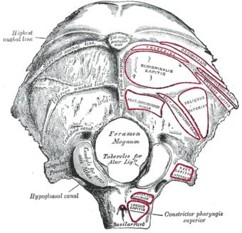
Occipital bone. Outer surface (red circle at upper right is for occipitalis)
|
|
| Details | |
| Latin | Venter occipitalis musculi occipitofrontalis |
| Origin | Superior nuchal line of the occipital bone and mastoid process of the temporal bone |
| Insertion | Galea aponeurosis |
| Occipital artery | |
| Posterior auricular nerve (facial nerve) | |
| Actions | Moves the scalp back |
| Identifiers | |
| Dorlands /Elsevier |
m_22/12549942 |
| TA | Lua error in Module:Wikidata at line 744: attempt to index field 'wikibase' (a nil value). |
| TH | {{#property:P1694}} |
| TE | {{#property:P1693}} |
| FMA | {{#property:P1402}} |
| Anatomical terms of muscle
[[[d:Lua error in Module:Wikidata at line 863: attempt to index field 'wikibase' (a nil value).|edit on Wikidata]]]
|
|
The occipitalis muscle (occipital belly) is a muscle which covers parts of the skull. Some sources consider the occipital muscle to be a distinct muscle. However, Terminologia Anatomica currently classifies it as part of the occipitofrontalis muscle along with the frontalis muscle.
The occipitalis muscle is thin and quadrilateral in form. It arises from tendinous fibers from the lateral two-thirds of the superior nuchal line of the occipital bone and from the mastoid process of the temporal and ends in the galea aponeurotica.[1]
The occipitalis muscle is innervated by the facial nerve and its function is to move the scalp back.[2] The muscles receives blood from the occipital artery.
Additional image
See also
References
This article incorporates text in the public domain from the 20th edition of Gray's Anatomy (1918)
